


Crystal structure of Putative membrane anchored protein from Corynebacterium diphtheriae
Chang, C., Sather, A., Clancy, S., Joachimiak, A.To be published.
Experimental Data Snapshot
wwPDB Validation 3D Report Full Report

Entity ID: 1 | |||||
|---|---|---|---|---|---|
| Molecule | Chains | Sequence Length | Organism | Details | Image |
| Putative membrane anchored protein | 489 | Corynebacterium diphtheriae | Mutation(s): 0 Gene Names: DIP2116 | 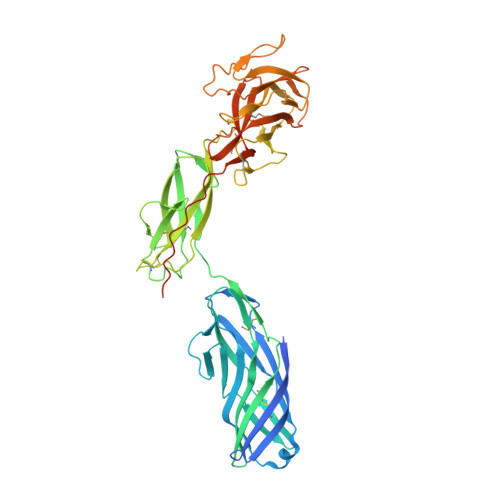 | |
UniProt | |||||
Find proteins for Q6NEZ3 (Corynebacterium diphtheriae (strain ATCC 700971 / NCTC 13129 / Biotype gravis)) Explore Q6NEZ3 Go to UniProtKB: Q6NEZ3 | |||||
Entity Groups | |||||
| Sequence Clusters | 30% Identity50% Identity70% Identity90% Identity95% Identity100% Identity | ||||
| UniProt Group | Q6NEZ3 | ||||
Sequence AnnotationsExpand | |||||
| |||||
Entity ID: 2 | |||||
|---|---|---|---|---|---|
| Molecule | Chains | Sequence Length | Organism | Details | Image |
| Putative membrane anchored protein | 489 | Corynebacterium diphtheriae | Mutation(s): 0 Gene Names: DIP2116 | 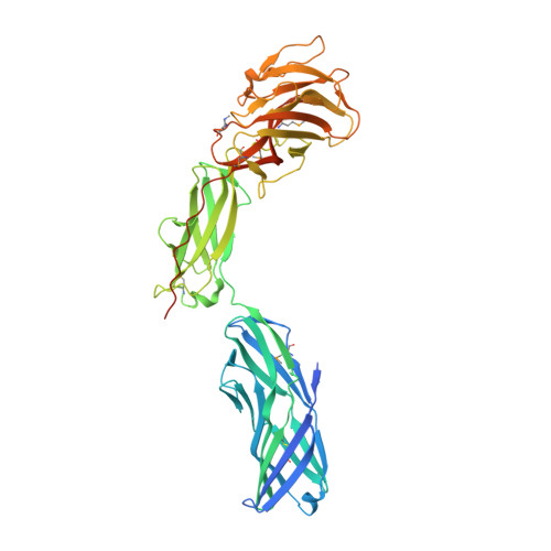 | |
UniProt | |||||
Find proteins for Q6NEZ3 (Corynebacterium diphtheriae (strain ATCC 700971 / NCTC 13129 / Biotype gravis)) Explore Q6NEZ3 Go to UniProtKB: Q6NEZ3 | |||||
Entity Groups | |||||
| Sequence Clusters | 30% Identity50% Identity70% Identity90% Identity95% Identity100% Identity | ||||
| UniProt Group | Q6NEZ3 | ||||
Sequence AnnotationsExpand | |||||
| |||||
| Ligands 1 Unique | |||||
|---|---|---|---|---|---|
| ID | Chains | Name / Formula / InChI Key | 2D Diagram | 3D Interactions | |
| CL Query on CL | C [auth B] | CHLORIDE ION Cl VEXZGXHMUGYJMC-UHFFFAOYSA-M |  | ||
| Modified Residues 2 Unique | |||||
|---|---|---|---|---|---|
| ID | Chains | Type | Formula | 2D Diagram | Parent |
| MLZ Query on MLZ | A | L-PEPTIDE LINKING | C7 H16 N2 O2 |  | LYS |
| MSE Query on MSE | A | L-PEPTIDE LINKING | C5 H11 N O2 Se |  | MET |
| Length ( Å ) | Angle ( ˚ ) |
|---|---|
| a = 73.205 | α = 90 |
| b = 95.517 | β = 90 |
| c = 160.778 | γ = 90 |
| Software Name | Purpose |
|---|---|
| SBC-Collect | data collection |
| HKL-3000 | phasing |
| SHELXD | phasing |
| SHELXE | model building |
| MLPHARE | phasing |
| DM | model building |
| RESOLVE | model building |
| Coot | model building |
| ARP/wARP | model building |
| REFMAC | refinement |
| HKL-3000 | data reduction |
| HKL-3000 | data scaling |
| DM | phasing |
| RESOLVE | phasing |
RCSB PDB (citation) is hosted by
RCSB PDB is a member of the

