3LQU

Crystal structure of 3,4-Dihydroxy-2-butanone 4-phosphate synthase complexed with Ribulose-5 phosphate


Experimental Data Snapshot

  • Method: X-RAY DIFFRACTION
  • Resolution: 2.52 Å
  • R-Value Free: 0.274 
  • R-Value Work: 0.182 
  • R-Value Observed: 0.186 

wwPDB Validation   3D Report Full Report


Ligand Structure Quality Assessment 


This is version 1.3 of the entry. See complete history


Literature

Potential anti-bacterial drug target: structural characterization of 3,4-dihydroxy-2-butanone-4-phosphate synthase from Salmonella typhimurium LT2.

Kumar, P.Singh, M.Gautam, R.Karthikeyan, S.

(2010) Proteins 78: 3292-3303

  • DOI: https://doi.org/10.1002/prot.22837
  • Primary Citation of Related Structures:  
    3LQU, 3LRJ, 3LS6

  • PubMed Abstract: 

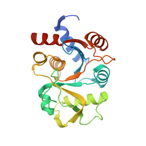
    3,4-Dihydroxy-2-butanone-4-phosphate synthase (DHBPS) encoded by ribB gene is one of the first enzymes in riboflavin biosynthesis pathway and catalyzes the conversion of ribulose-5-phosphate (Ru5P) to 3,4-dihydroxy-2-butanone-4-phosphate and formate. DHBPS is an attractive target for developing anti-bacterial drugs as this enzyme is essential for pathogens, but absent in humans. The recombinant DHBPS enzyme of Salmonella requires magnesium ion for its activity and catalyzes the formation of 3,4-dihydroxy-2-butanone-4-phosphate from Ru5P at a rate of 199 nmol min(-1) mg(-1) with K(m) value of 116 μM at 37°C. Further, we have determined the crystal structures of Salmonella DHBPS in complex with sulfate, Ru5P and sulfate-zinc ion at a resolution of 2.80, 2.52, and 1.86 Å, respectively. Analysis of these crystal structures reveals that the acidic loop (residues 34-39) responsible for the acid-base catalysis is disordered in the absence of substrate or metal ion at the active site. Upon binding either substrate or sulfate and metal ions, the acidic loop becomes stabilized, adopts a closed conformation and interacts with the substrate. Our structure for the first time reveals that binding of substrate Ru5P alone is sufficient for the stabilization of the acidic active site loop into a closed conformation. In addition, the Glu38 residue from the acidic active site loop undergoes a conformational change upon Ru5P binding, which helps in positioning the second metal ion that stabilizes the Ru5P and the reaction intermediates. This is the first structural report of DHBPS in complex with either substrate or metal ion from any eubacteria.


  • Organizational Affiliation

    Institute of Microbial Technology, Council of Scientific and Industrial Research (CSIR), Sector 39-A, Chandigarh 160 036, India.


Macromolecules
Find similar proteins by:  (by identity cutoff)  |  3D Structure
Entity ID: 1
MoleculeChains Sequence LengthOrganismDetailsImage
3,4-dihydroxy-2-butanone 4-phosphate synthase
A, B
217Salmonella enterica subsp. enterica serovar TyphimuriumMutation(s): 0 
Gene Names: ribBSTM3195
EC: 4.1.99.12
UniProt
Find proteins for P66032 (Salmonella typhimurium (strain LT2 / SGSC1412 / ATCC 700720))
Explore P66032 
Go to UniProtKB:  P66032
Entity Groups  
Sequence Clusters30% Identity50% Identity70% Identity90% Identity95% Identity100% Identity
UniProt GroupP66032
Sequence Annotations
Expand
  • Reference Sequence
Small Molecules
Ligands 1 Unique
IDChains Name / Formula / InChI Key2D Diagram3D Interactions
5SP
Query on 5SP

Download Ideal Coordinates CCD File 
C [auth A]5-O-phosphono-D-xylulose
C5 H11 O8 P
FNZLKVNUWIIPSJ-RFZPGFLSSA-N
Experimental Data & Validation

Experimental Data

  • Method: X-RAY DIFFRACTION
  • Resolution: 2.52 Å
  • R-Value Free: 0.274 
  • R-Value Work: 0.182 
  • R-Value Observed: 0.186 
  • Space Group: C 2 2 21
Unit Cell:
Length ( Å )Angle ( ˚ )
a = 66.236α = 90
b = 77.386β = 90
c = 149.372γ = 90
Software Package:
Software NamePurpose
MAR345dtbdata collection
PHASERphasing
CCP4model building
PHENIXrefinement
HKL-2000data reduction
HKL-2000data scaling
CCP4phasing

Structure Validation

View Full Validation Report


Ligand Structure Quality Assessment 


Entry History 

Deposition Data

Revision History  (Full details and data files)

  • Version 1.0: 2010-09-15
    Type: Initial release
  • Version 1.1: 2011-07-13
    Changes: Version format compliance
  • Version 1.2: 2014-03-05
    Changes: Database references
  • Version 1.3: 2023-11-01
    Changes: Data collection, Database references, Derived calculations, Refinement description