3LQC

X-ray crystal structure of oxidized XRCC1 bound to DNA pol beta Palm thumb domain


Experimental Data Snapshot

  • Method: X-RAY DIFFRACTION
  • Resolution: 2.35 Å
  • R-Value Free: 0.250 
  • R-Value Work: 0.189 
  • R-Value Observed: 0.192 

wwPDB Validation   3D Report Full Report


This is version 1.2 of the entry. See complete history


Literature

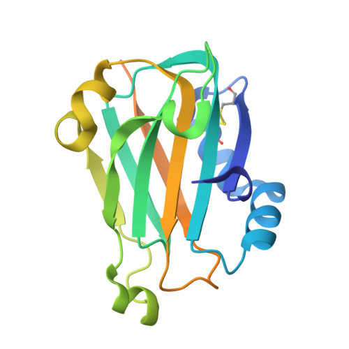
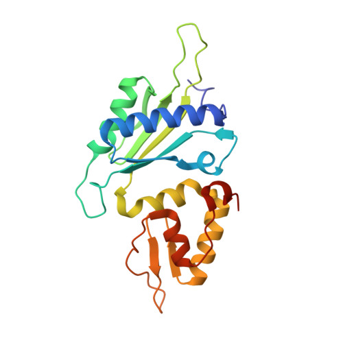
Oxidation state of the XRCC1 N-terminal domain regulates DNA polymerase beta binding affinity.

Cuneo, M.J.London, R.E.

(2010) Proc Natl Acad Sci U S A 107: 6805-6810

  • DOI: https://doi.org/10.1073/pnas.0914077107
  • Primary Citation of Related Structures:  
    3K75, 3K77, 3LQC

  • PubMed Abstract: 

    Formation of a complex between the XRCC1 N-terminal domain (NTD) and DNA polymerase beta (Pol beta) is central to base excision repair of damaged DNA. Two crystal forms of XRCC1-NTD complexed with Pol beta have been solved, revealing that the XRCC1-NTD is able to adopt a redox-dependent alternate fold, characterized by a disulfide bond, and substantial variations of secondary structure, folding topology, and electrostatic surface. Although most of these structural changes occur distal to the interface, the oxidized XRCC1-NTD forms additional interactions with Pol beta, enhancing affinity by an order of magnitude. Transient disulfide bond formation is increasingly recognized as an important molecular regulatory mechanism. The results presented here suggest a paradigm in DNA repair in which the redox state of a scaffolding protein plays an active role in organizing the repair complex.


  • Organizational Affiliation

    Laboratory of Structural Biology, National Institute of Environmental Health Sciences, Research Triangle Park, NC 27709, USA.


Macromolecules
Find similar proteins by:  (by identity cutoff)  |  3D Structure
Entity ID: 1
MoleculeChains Sequence LengthOrganismDetailsImage
DNA repair protein XRCC1189Homo sapiensMutation(s): 0 
Gene Names: XRCC1
UniProt & NIH Common Fund Data Resources
Find proteins for P18887 (Homo sapiens)
Explore P18887 
Go to UniProtKB:  P18887
PHAROS:  P18887
GTEx:  ENSG00000073050 
Entity Groups  
Sequence Clusters30% Identity50% Identity70% Identity90% Identity95% Identity100% Identity
UniProt GroupP18887
Sequence Annotations
Expand
  • Reference Sequence
Find similar proteins by:  (by identity cutoff)  |  3D Structure
Entity ID: 2
MoleculeChains Sequence LengthOrganismDetailsImage
DNA polymerase beta200Rattus norvegicusMutation(s): 0 
Gene Names: Polb
EC: 2.7.7.7 (PDB Primary Data), 4.2.99 (PDB Primary Data)
UniProt
Find proteins for P06766 (Rattus norvegicus)
Explore P06766 
Go to UniProtKB:  P06766
Entity Groups  
Sequence Clusters30% Identity50% Identity70% Identity90% Identity95% Identity100% Identity
UniProt GroupP06766
Sequence Annotations
Expand
  • Reference Sequence
Experimental Data & Validation

Experimental Data

  • Method: X-RAY DIFFRACTION
  • Resolution: 2.35 Å
  • R-Value Free: 0.250 
  • R-Value Work: 0.189 
  • R-Value Observed: 0.192 
  • Space Group: P 31 2 1
Unit Cell:
Length ( Å )Angle ( ˚ )
a = 75.24α = 90
b = 75.24β = 90
c = 126.16γ = 120
Software Package:
Software NamePurpose
XSCALEdata scaling
PHENIXrefinement
PDB_EXTRACTdata extraction
HKL-2000data collection
PHASERphasing

Structure Validation

View Full Validation Report


Entry History 

Deposition Data

Revision History  (Full details and data files)

  • Version 1.0: 2010-04-28
    Type: Initial release
  • Version 1.1: 2011-07-13
    Changes: Version format compliance
  • Version 1.2: 2023-09-06
    Changes: Data collection, Database references, Derived calculations, Refinement description, Structure summary