Proenzyme structure and activation of astacin metallopeptidase
Guevara, T., Yiallouros, I., Kappelhoff, R., Bissdorf, S., Stocker, W., Gomis-Ruth, F.X.(2010) J Biol Chem 285: 13958-13965
- PubMed: 20202938
- DOI: https://doi.org/10.1074/jbc.M109.097436
- Primary Citation of Related Structures:
3LQ0 - PubMed Abstract:
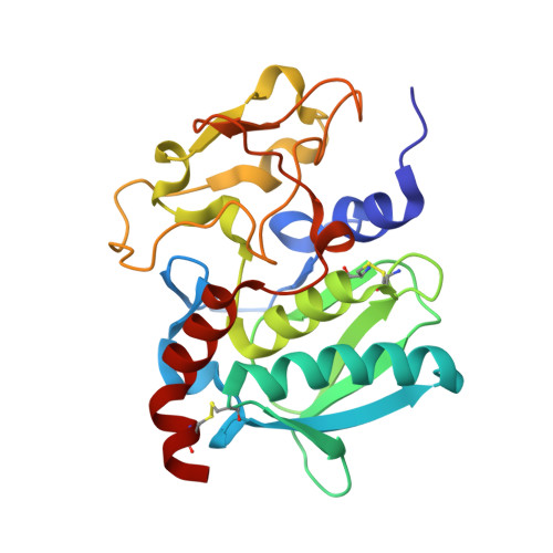
Proteolysis is regulated by inactive (latent) zymogens, with a prosegment preventing access of substrates to the active-site cleft of the enzyme. How latency is maintained often depends on the catalytic mechanism of the protease. For example, in several families of the metzincin metallopeptidases, a "cysteine switch" mechanism involves a conserved prosegment motif with a cysteine residue that coordinates the catalytic zinc ion. Another family of metzincins, the astacins, do not possess a cysteine switch, so latency is maintained by other means. We have solved the high resolution crystal structure of proastacin from the European crayfish, Astacus astacus. Its prosegment is the shortest structurally reported for a metallopeptidase, and it has a unique structure. It runs through the active-site cleft in reverse orientation to a genuine substrate. Moreover, a conserved aspartate, projected by a wide loop of the prosegment, coordinates the zinc ion instead of the catalytic solvent molecule found in the mature enzyme. Activation occurs through two-step limited proteolysis and entails major rearrangement of a flexible activation domain, which becomes rigid and creates the base of the substrate-binding cleft. Maturation also requires the newly formed N terminus to be precisely trimmed so that it can participate in a buried solvent-mediated hydrogen-bonding network, which includes an invariant active-site residue. We describe a novel mechanism for latency and activation, which shares some common features both with other metallopeptidases and with serine peptidases.
Organizational Affiliation:
Proteolysis Laboratory, Department of Structural Biology, Molecular Biology Institute of Barcelona, Consejo Superior de Investigaciones Científicas, Barcelona Science Park, Helix Building, c/Baldiri Reixac 15-21, E-08028, Spain.


