3LPH

Crystal structure of the HIV-1 Rev dimer


Experimental Data Snapshot

  • Method: X-RAY DIFFRACTION
  • Resolution: 2.50 Å
  • R-Value Free: 0.261 
  • R-Value Work: 0.226 
  • R-Value Observed: 0.228 

wwPDB Validation   3D Report Full Report


This is version 1.5 of the entry. See complete history


Literature

Structural basis for cooperative RNA binding and export complex assembly by HIV Rev.

Daugherty, M.D.Liu, B.Frankel, A.D.

(2010) Nat Struct Mol Biol 17: 1337-1342

  • DOI: https://doi.org/10.1038/nsmb.1902
  • Primary Citation of Related Structures:  
    3LPH

  • PubMed Abstract: 

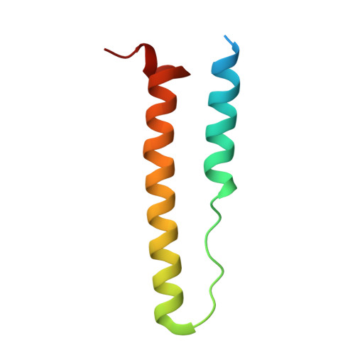
    HIV replication requires nuclear export of unspliced viral RNAs to translate structural proteins and package genomic RNA. Export is mediated by cooperative binding of the Rev protein to the Rev response element (RRE) RNA, to form a highly specific oligomeric ribonucleoprotein (RNP) that binds to the Crm1 host export factor. To understand how protein oligomerization generates cooperativity and specificity for RRE binding, we solved the crystal structure of a Rev dimer at 2.5-Å resolution. The dimer arrangement organizes arginine-rich helices at the ends of a V-shaped assembly to bind adjacent RNA sites and structurally couple dimerization and RNA recognition. A second protein-protein interface arranges higher-order Rev oligomers to act as an adaptor to the host export machinery, with viral RNA bound to one face and Crm1 to another, the oligomers thereby using small, interconnected modules to physically arrange the RNP for efficient export.


  • Organizational Affiliation

    Chemistry and Chemical Biology Graduate Program, University of California, San Francisco, San Francisco, California, USA.


Macromolecules
Find similar proteins by:  (by identity cutoff)  |  3D Structure
Entity ID: 1
MoleculeChains Sequence LengthOrganismDetailsImage
Protein Rev
A, B, C, D
72Human immunodeficiency virus type 1 (HXB3 ISOLATE)Mutation(s): 2 
Gene Names: rev
UniProt
Find proteins for P69718 (Human immunodeficiency virus type 1 group M subtype B (isolate HXB3))
Explore P69718 
Go to UniProtKB:  P69718
Entity Groups  
Sequence Clusters30% Identity50% Identity70% Identity90% Identity95% Identity100% Identity
UniProt GroupP69718
Sequence Annotations
Expand
  • Reference Sequence
Small Molecules
Ligands 3 Unique
IDChains Name / Formula / InChI Key2D Diagram3D Interactions
MLI
Query on MLI

Download Ideal Coordinates CCD File 
J [auth A],
O [auth B]
MALONATE ION
C3 H2 O4
OFOBLEOULBTSOW-UHFFFAOYSA-L
SO4
Query on SO4

Download Ideal Coordinates CCD File 
E [auth A],
F [auth A]
SULFATE ION
O4 S
QAOWNCQODCNURD-UHFFFAOYSA-L
BR
Query on BR

Download Ideal Coordinates CCD File 
G [auth A]
H [auth A]
I [auth A]
K [auth B]
L [auth B]
G [auth A],
H [auth A],
I [auth A],
K [auth B],
L [auth B],
M [auth B],
N [auth B],
P [auth C],
Q [auth C],
R [auth C],
S [auth C],
T [auth D],
U [auth D]
BROMIDE ION
Br
CPELXLSAUQHCOX-UHFFFAOYSA-M
Experimental Data & Validation

Experimental Data

  • Method: X-RAY DIFFRACTION
  • Resolution: 2.50 Å
  • R-Value Free: 0.261 
  • R-Value Work: 0.226 
  • R-Value Observed: 0.228 
  • Space Group: P 64 2 2
Unit Cell:
Length ( Å )Angle ( ˚ )
a = 115.801α = 90
b = 115.801β = 90
c = 81.163γ = 120
Software Package:
Software NamePurpose
DENZOdata reduction
SCALEPACKdata scaling
SOLVEphasing
RESOLVEphasing
PHENIXrefinement
PDB_EXTRACTdata extraction
Blu-Icedata collection
HKL-2000data reduction
HKL-2000data scaling
PHENIXphasing

Structure Validation

View Full Validation Report


Entry History 

Deposition Data

Revision History  (Full details and data files)

  • Version 1.0: 2010-12-08
    Type: Initial release
  • Version 1.1: 2011-07-13
    Changes: Version format compliance
  • Version 1.2: 2017-11-01
    Changes: Refinement description
  • Version 1.3: 2021-10-13
    Changes: Database references, Derived calculations
  • Version 1.4: 2024-02-21
    Changes: Data collection
  • Version 1.5: 2024-04-03
    Changes: Refinement description