


Crystal structure of guanylate kinase from Anaplasma phagocytophilum
Abendroth, J., Sankaran, B., Davies, D., Staker, B.To be published.
Experimental Data Snapshot
Entity ID: 1 | |||||
|---|---|---|---|---|---|
| Molecule | Chains | Sequence Length | Organism | Details | Image |
| Guanylate kinase | 231 | Anaplasma phagocytophilum str. HZ | Mutation(s): 0 Gene Names: gmk, APH_0170 EC: 2.7.4.8 | 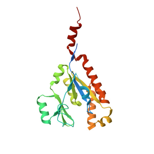 | |
UniProt | |||||
Find proteins for Q2GLF7 (Anaplasma phagocytophilum (strain HZ)) Explore Q2GLF7 Go to UniProtKB: Q2GLF7 | |||||
Entity Groups | |||||
| Sequence Clusters | 30% Identity50% Identity70% Identity90% Identity95% Identity100% Identity | ||||
| UniProt Group | Q2GLF7 | ||||
Sequence AnnotationsExpand | |||||
| |||||
| Ligands 1 Unique | |||||
|---|---|---|---|---|---|
| ID | Chains | Name / Formula / InChI Key | 2D Diagram | 3D Interactions | |
| 5GP Query on 5GP | C [auth A], D [auth B] | GUANOSINE-5'-MONOPHOSPHATE C10 H14 N5 O8 P RQFCJASXJCIDSX-UUOKFMHZSA-N |  | ||
| Length ( Å ) | Angle ( ˚ ) |
|---|---|
| a = 64.53 | α = 90 |
| b = 76.58 | β = 90 |
| c = 103.66 | γ = 90 |
| Software Name | Purpose |
|---|---|
| BOS | data collection |
| PHASER | phasing |
| REFMAC | refinement |
| XDS | data reduction |
| XSCALE | data scaling |
RCSB PDB (citation) is hosted by
RCSB PDB is a member of the

