


The crystal structure of the protein NE1376 with unknown function from Nitrosomonas europaea ATCC 19718
Zhang, R., Kagan, O., Savchenko, A., Joachimiak, A., Edwards, A.To be published.
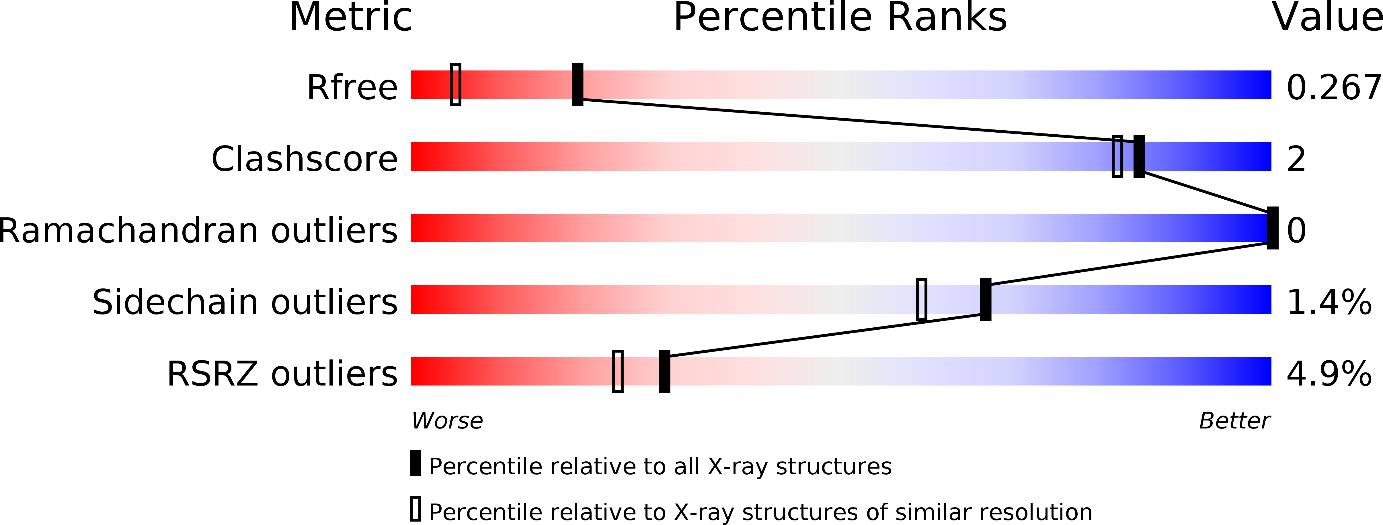
Experimental Data Snapshot
wwPDB Validation 3D Report Full Report
Entity ID: 1 | |||||
|---|---|---|---|---|---|
| Molecule | Chains | Sequence Length | Organism | Details | Image |
| Uncharacterized protein | 83 | Pseudomonas aeruginosa | Mutation(s): 0 Gene Names: GI:15599178, PA3983 | 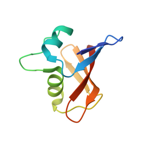 | |
UniProt | |||||
Find proteins for Q9HX36 (Pseudomonas aeruginosa (strain ATCC 15692 / DSM 22644 / CIP 104116 / JCM 14847 / LMG 12228 / 1C / PRS 101 / PAO1)) Explore Q9HX36 Go to UniProtKB: Q9HX36 | |||||
Entity Groups | |||||
| Sequence Clusters | 30% Identity50% Identity70% Identity90% Identity95% Identity100% Identity | ||||
| UniProt Group | Q9HX36 | ||||
Sequence AnnotationsExpand | |||||
| |||||
| Length ( Å ) | Angle ( ˚ ) |
|---|---|
| a = 73.034 | α = 90 |
| b = 73.034 | β = 90 |
| c = 30.728 | γ = 90 |
| Software Name | Purpose |
|---|---|
| SBC-Collect | data collection |
| HKL-3000 | phasing |
| REFMAC | refinement |
| HKL-3000 | data reduction |
| HKL-3000 | data scaling |
RCSB PDB (citation) is hosted by
RCSB PDB is a member of the

