3LKA

Catalytic domain of human MMP-12 complexed with hydroxamic acid and paramethoxy-sulfonyl amide


Experimental Data Snapshot

  • Method: X-RAY DIFFRACTION
  • Resolution: 1.80 Å
  • R-Value Free: 0.210 
  • R-Value Work: 0.169 
  • R-Value Observed: 0.173 

wwPDB Validation   3D Report Full Report


Ligand Structure Quality Assessment 


This is version 1.4 of the entry. See complete history


Literature

Entropic contribution to the linking coefficient in fragment based drug design: a case study.

Borsi, V.Calderone, V.Fragai, M.Luchinat, C.Sarti, N.

(2010) J Med Chem 53: 4285-4289

  • DOI: https://doi.org/10.1021/jm901723z
  • Primary Citation of Related Structures:  
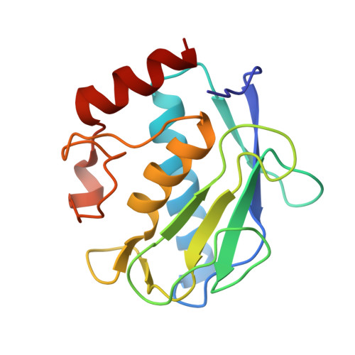
    3LKA

  • PubMed Abstract: 

    For several drug leads obtained by tethering weak binding ligands, the dissociation constant is smaller than the product of those of the individual fragments by a factor named the linking coefficient, E. This favorable contribution is attributed to the entropic gain that is realized when two weak binding ligands are tethered. Here we show a case study where the linking coefficient is strikingly small (E = 2.1 x 10(-3) M(-1)) and its totally entropic nature is demonstrated.


  • Organizational Affiliation

    Magnetic Resonance Center (CERM), University of Florence, Florence, Italy.


Macromolecules
Find similar proteins by:  (by identity cutoff)  |  3D Structure
Entity ID: 1
MoleculeChains Sequence LengthOrganismDetailsImage
Macrophage metalloelastase158Homo sapiensMutation(s): 1 
Gene Names: HMEMMP12
EC: 3.4.24.65
UniProt & NIH Common Fund Data Resources
Find proteins for P39900 (Homo sapiens)
Explore P39900 
Go to UniProtKB:  P39900
PHAROS:  P39900
GTEx:  ENSG00000262406 
Entity Groups  
Sequence Clusters30% Identity50% Identity70% Identity90% Identity95% Identity100% Identity
UniProt GroupP39900
Sequence Annotations
Expand
  • Reference Sequence
Small Molecules
Ligands 4 Unique
IDChains Name / Formula / InChI Key2D Diagram3D Interactions
M4S
Query on M4S

Download Ideal Coordinates CCD File 
H [auth A]4-methoxybenzenesulfonamide
C7 H9 N O3 S
MSFQEZBRFPAFEX-UHFFFAOYSA-N
HAE
Query on HAE

Download Ideal Coordinates CCD File 
G [auth A]ACETOHYDROXAMIC ACID
C2 H5 N O2
RRUDCFGSUDOHDG-UHFFFAOYSA-N
ZN
Query on ZN

Download Ideal Coordinates CCD File 
B [auth A],
C [auth A]
ZINC ION
Zn
PTFCDOFLOPIGGS-UHFFFAOYSA-N
CA
Query on CA

Download Ideal Coordinates CCD File 
D [auth A],
E [auth A],
F [auth A]
CALCIUM ION
Ca
BHPQYMZQTOCNFJ-UHFFFAOYSA-N
Binding Affinity Annotations 
IDSourceBinding Affinity
M4S PDBBind:  3LKA Kd: 1.50e+6 (nM) from 1 assay(s)
Binding MOAD:  3LKA Kd: 1.50e+6 (nM) from 1 assay(s)
Experimental Data & Validation

Experimental Data

  • Method: X-RAY DIFFRACTION
  • Resolution: 1.80 Å
  • R-Value Free: 0.210 
  • R-Value Work: 0.169 
  • R-Value Observed: 0.173 
  • Space Group: C 1 2 1
Unit Cell:
Length ( Å )Angle ( ˚ )
a = 51.566α = 90
b = 60.194β = 115.09
c = 54.215γ = 90
Software Package:
Software NamePurpose
CrysalisProdata collection
MOLREPphasing
REFMACrefinement
MOSFLMdata reduction
SCALAdata scaling

Structure Validation

View Full Validation Report


Ligand Structure Quality Assessment 


Entry History 

Deposition Data

Revision History  (Full details and data files)

  • Version 1.0: 2010-05-12
    Type: Initial release
  • Version 1.1: 2011-07-13
    Changes: Version format compliance
  • Version 1.2: 2017-11-01
    Changes: Refinement description
  • Version 1.3: 2021-10-13
    Changes: Database references, Derived calculations
  • Version 1.4: 2023-09-06
    Changes: Data collection, Refinement description