3LJ2

IRE1 complexed with JAK Inhibitor I


Experimental Data Snapshot

  • Method: X-RAY DIFFRACTION
  • Resolution: 3.33 Å
  • R-Value Free: 0.293 
  • R-Value Work: 0.254 
  • R-Value Observed: 0.256 

wwPDB Validation   3D Report Full Report


Ligand Structure Quality Assessment 


This is version 1.3 of the entry. See complete history


Literature

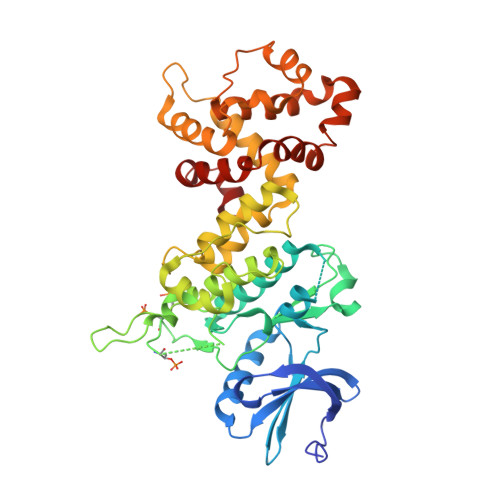
Flavonol activation defines an unanticipated ligand-binding site in the kinase-RNase domain of IRE1.

Wiseman, R.L.Zhang, Y.Lee, K.P.Harding, H.P.Haynes, C.M.Price, J.Sicheri, F.Ron, D.

(2010) Mol Cell 38: 291-304

  • DOI: https://doi.org/10.1016/j.molcel.2010.04.001
  • Primary Citation of Related Structures:  
    3LJ0, 3LJ1, 3LJ2

  • PubMed Abstract: 

    Signaling in the most conserved branch of the endoplasmic reticulum (ER) unfolded protein response (UPR) is initiated by sequence-specific cleavage of the HAC1/XBP1 mRNA by the ER stress-induced kinase-endonuclease IRE1. We have discovered that the flavonol quercetin activates yeast IRE1's RNase and potentiates activation by ADP, a natural activating ligand that engages the IRE1 nucleotide-binding cleft. Enzyme kinetics and the structure of a cocrystal of IRE1 complexed with ADP and quercetin reveal engagement by quercetin of an unanticipated ligand-binding pocket at the dimer interface of IRE1's kinase extension nuclease (KEN) domain. Analytical ultracentrifugation and crosslinking studies support the preeminence of enhanced dimer formation in quercetin's mechanism of action. These findings hint at the existence of endogenous cytoplasmic ligands that may function alongside stress signals from the ER lumen to modulate IRE1 activity and at the potential for the development of drugs that modify UPR signaling from this unanticipated site.


  • Organizational Affiliation

    Kimmel Center for Biology and Medicine of the Skirball Institute, New York University School of Medicine, New York, NY 10016, USA.


Macromolecules
Find similar proteins by:  (by identity cutoff)  |  3D Structure
Entity ID: 1
MoleculeChains Sequence LengthOrganismDetailsImage
Serine/threonine-protein kinase/endoribonuclease IRE1
A, B
434Saccharomyces cerevisiaeMutation(s): 0 
Gene Names: ERN1IRE1YHR079C
EC: 2.7.11.1 (PDB Primary Data), 3.1.26 (PDB Primary Data)
UniProt
Find proteins for P32361 (Saccharomyces cerevisiae (strain ATCC 204508 / S288c))
Explore P32361 
Go to UniProtKB:  P32361
Entity Groups  
Sequence Clusters30% Identity50% Identity70% Identity90% Identity95% Identity100% Identity
UniProt GroupP32361
Sequence Annotations
Expand
  • Reference Sequence
Small Molecules
Ligands 1 Unique
IDChains Name / Formula / InChI Key2D Diagram3D Interactions
IZA
Query on IZA

Download Ideal Coordinates CCD File 
C [auth A],
D [auth B]
2-TERT-BUTYL-9-FLUORO-3,6-DIHYDRO-7H-BENZ[H]-IMIDAZ[4,5-F]ISOQUINOLINE-7-ONE
C18 H16 F N3 O
VNDWQCSOSCCWIP-UHFFFAOYSA-N
Modified Residues  2 Unique
IDChains TypeFormula2D DiagramParent
SEP
Query on SEP
A, B
L-PEPTIDE LINKINGC3 H8 N O6 PSER
TPO
Query on TPO
A, B
L-PEPTIDE LINKINGC4 H10 N O6 PTHR
Experimental Data & Validation

Experimental Data

  • Method: X-RAY DIFFRACTION
  • Resolution: 3.33 Å
  • R-Value Free: 0.293 
  • R-Value Work: 0.254 
  • R-Value Observed: 0.256 
  • Space Group: P 32 1 2
Unit Cell:
Length ( Å )Angle ( ˚ )
a = 126.18α = 90
b = 126.18β = 90
c = 175.16γ = 120
Software Package:
Software NamePurpose
SCALAdata scaling
REFMACrefinement
PDB_EXTRACTdata extraction
MOSFLMdata reduction
PHASERphasing

Structure Validation

View Full Validation Report


Ligand Structure Quality Assessment 


Entry History 

Deposition Data

Revision History  (Full details and data files)

  • Version 1.0: 2010-05-12
    Type: Initial release
  • Version 1.1: 2011-07-13
    Changes: Advisory, Refinement description, Version format compliance
  • Version 1.2: 2017-11-01
    Changes: Refinement description
  • Version 1.3: 2023-09-06
    Changes: Data collection, Database references, Derived calculations, Refinement description