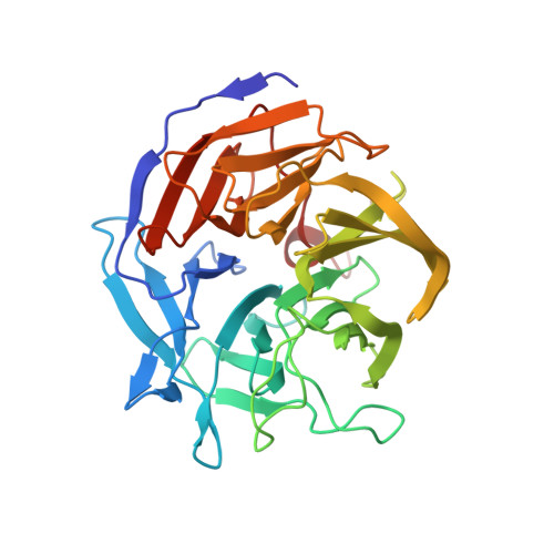
Structural characterization of the catalytic calcium-binding site in diisopropyl fluorophosphatase (DFPase)-Comparison with related beta-propeller enzymes.
Blum, M.M., Chen, J.C.(2010) Chem Biol Interact 187: 373-379
- PubMed: 20206152
- DOI: https://doi.org/10.1016/j.cbi.2010.02.043
- Primary Citation of Related Structures:
3LI3, 3LI4, 3LI5 - PubMed Abstract:
The calcium-dependent phosphotriesterase diisopropyl fluorophosphatase (DFPase) from the squid Loligo vulgaris efficiently hydrolyzes a wide range of organophosphorus nerve agents. The two calcium ions within DFPase play essential roles for its function. The lower affinity calcium ion located at the bottom of the active site participates in the reaction mechanism, while the high affinity calcium in the center of the protein maintains structural integrity of the enzyme. The activity and structures of three DFPase variants targeting the catalytic calcium-binding site are reported (D121E, N120D/N175D/D229N, and E21Q/N120D/N175D/D229N), and the effect of these mutations on the overall structural dynamics of DFPase is examined using molecular dynamics simulations. While D229 is crucial for enzymatic activity, E21 is essential for calcium binding. Although at least two negatively charged side chains are required for calcium binding, the addition of a third charge significantly lowers the activity. Furthermore, the arrangement of these charges in the binding site is important for enzymatic activity. These results, together with earlier mutational, structural, and kinetic studies, show a highly evolved calcium-binding environment, with a specific electrostatic topology crucial for activity. A number of structural homologues of DFPase have been recently identified, including a chimeric variant of Paraoxonase 1 (PON1), drug resistance protein 35 (Drp35) from Staphylococcus aureus and the gluconolactonase XC5397 from Xanthomonas campestris. Surprisingly, despite low sequence identity, these proteins share remarkably similar calcium-binding environments to DFPase.
Organizational Affiliation:
Blum-Scientific Services, Ledererstrasse 23, 80331 Munich, Germany. mmblum@blum-scientific.de