


Crystal structure of a putative agmatinase from Clostridium difficile
Palani, K., Burley, S.K., Swaminathan, S.To be published.
Experimental Data Snapshot
wwPDB Validation 3D Report Full Report

Entity ID: 1 | |||||
|---|---|---|---|---|---|
| Molecule | Chains | Sequence Length | Organism | Details | Image |
| Putative agmatinase | 287 | Clostridioides difficile 630 | Mutation(s): 0 Gene Names: CD0891, speB | 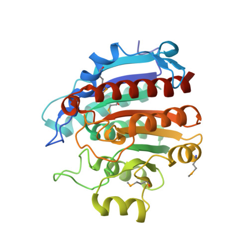 | |
UniProt | |||||
Find proteins for Q18A84 (Clostridioides difficile (strain 630)) Explore Q18A84 Go to UniProtKB: Q18A84 | |||||
Entity Groups | |||||
| Sequence Clusters | 30% Identity50% Identity70% Identity90% Identity95% Identity100% Identity | ||||
| UniProt Group | Q18A84 | ||||
Sequence AnnotationsExpand | |||||
| |||||
| Ligands 3 Unique | |||||
|---|---|---|---|---|---|
| ID | Chains | Name / Formula / InChI Key | 2D Diagram | 3D Interactions | |
| MPD Query on MPD | H [auth A] | (4S)-2-METHYL-2,4-PENTANEDIOL C6 H14 O2 SVTBMSDMJJWYQN-YFKPBYRVSA-N |  | ||
| PO4 Query on PO4 | F [auth A], G [auth A] | PHOSPHATE ION O4 P NBIIXXVUZAFLBC-UHFFFAOYSA-K |  | ||
| MN Query on MN | B [auth A], C [auth A], D [auth A], E [auth A] | MANGANESE (II) ION Mn WAEMQWOKJMHJLA-UHFFFAOYSA-N |  | ||
| Modified Residues 1 Unique | |||||
|---|---|---|---|---|---|
| ID | Chains | Type | Formula | 2D Diagram | Parent |
| MSE Query on MSE | A | L-PEPTIDE LINKING | C5 H11 N O2 Se |  | MET |
| Length ( Å ) | Angle ( ˚ ) |
|---|---|
| a = 77.35 | α = 90 |
| b = 77.35 | β = 90 |
| c = 166.118 | γ = 120 |
| Software Name | Purpose |
|---|---|
| CNS | refinement |
| CBASS | data collection |
| HKL-2000 | data reduction |
| HKL-2000 | data scaling |
| SHELXD | phasing |
| SHARP | phasing |
| ARP/wARP | model building |
RCSB PDB (citation) is hosted by
RCSB PDB is a member of the

