3LGS

A. thaliana MTA nucleosidase in complex with S-adenosylhomocysteine


Experimental Data Snapshot

  • Method: X-RAY DIFFRACTION
  • Resolution: 2.20 Å
  • R-Value Free: 0.260 
  • R-Value Work: 0.210 
  • R-Value Observed: 0.210 

wwPDB Validation   3D Report Full Report


Ligand Structure Quality Assessment 


This is version 1.2 of the entry. See complete history


Literature

Mechanism of substrate specificity in 5'-methylthioadenosine/S-adenosylhomocysteine nucleosidases.

Siu, K.K.Asmus, K.Zhang, A.N.Horvatin, C.Li, S.Liu, T.Moffatt, B.Woods, V.L.Howell, P.L.

(2011) J Struct Biol 173: 86-98

  • DOI: https://doi.org/10.1016/j.jsb.2010.06.006
  • Primary Citation of Related Structures:  
    3LGS

  • PubMed Abstract: 

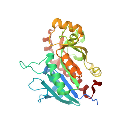
    5'-Methylthioadenosine/S-adenosylhomocysteine (MTA/SAH) nucleosidase (MTAN) plays a key role in the methionine-recycling pathway of bacteria and plants. Despite extensive structural and biochemical studies, the molecular mechanism of substrate specificity for MTAN remains an outstanding question. Bacterial MTANs show comparable efficiency in hydrolyzing MTA and SAH, while the plant enzymes select preferentially for MTA, with either no or significantly reduced activity towards SAH. Bacterial and plant MTANs show significant conservation in the overall structure, and the adenine- and ribose-binding sites. The observation of a more constricted 5'-alkylthio binding site in Arabidopsis thalianaAtMTAN1 and AtMTAN2, two plant MTAN homologues, led to the hypothesis that steric hindrance may play a role in substrate selection in plant MTANs. We show using isothermal titration calorimetry that SAH binds to both Escherichia coli MTAN (EcMTAN) and AtMTAN1 with comparable micromolar affinity. To understand why AtMTAN1 can bind but not hydrolyze SAH, we determined the structure of the protein-SAH complex at 2.2Å resolution. The lack of catalytic activity appears to be related to the enzyme's inability to bind the substrate in a catalytically competent manner. The role of dynamics in substrate selection was also examined by probing the amide proton exchange rates of EcMTAN and AtMTAN1 via deuterium-hydrogen exchange coupled mass spectrometry. These results correlate with the B factors of available structures and the thermodynamic parameters associated with substrate binding, and suggest a higher level of conformational flexibility in the active site of EcMTAN. Our results implicate dynamics as an important factor in substrate selection in MTAN.


  • Organizational Affiliation

    Research Institute, The Hospital for Sick Children, 555 University Avenue, Toronto, Ontario, Canada.


Macromolecules
Find similar proteins by:  (by identity cutoff)  |  3D Structure
Entity ID: 1
MoleculeChains Sequence LengthOrganismDetailsImage
5'-methylthioadenosine nucleosidases
A, B, C, D
267Arabidopsis thalianaMutation(s): 0 
Gene Names: AT4g38800atmtan1
EC: 3.2.2.16
UniProt
Find proteins for Q9T0I8 (Arabidopsis thaliana)
Explore Q9T0I8 
Go to UniProtKB:  Q9T0I8
Entity Groups  
Sequence Clusters30% Identity50% Identity70% Identity90% Identity95% Identity100% Identity
UniProt GroupQ9T0I8
Sequence Annotations
Expand
  • Reference Sequence
Small Molecules
Ligands 3 Unique
IDChains Name / Formula / InChI Key2D Diagram3D Interactions
SAH
Query on SAH

Download Ideal Coordinates CCD File 
E [auth A],
I [auth B],
N [auth C],
Q [auth D]
S-ADENOSYL-L-HOMOCYSTEINE
C14 H20 N6 O5 S
ZJUKTBDSGOFHSH-WFMPWKQPSA-N
ADE
Query on ADE

Download Ideal Coordinates CCD File 
F [auth A],
J [auth B],
O [auth C],
R [auth D]
ADENINE
C5 H5 N5
GFFGJBXGBJISGV-UHFFFAOYSA-N
EDO
Query on EDO

Download Ideal Coordinates CCD File 
G [auth A]
H [auth A]
K [auth B]
L [auth B]
M [auth B]
G [auth A],
H [auth A],
K [auth B],
L [auth B],
M [auth B],
P [auth C],
S [auth D],
T [auth D],
U [auth D]
1,2-ETHANEDIOL
C2 H6 O2
LYCAIKOWRPUZTN-UHFFFAOYSA-N
Binding Affinity Annotations 
IDSourceBinding Affinity
SAH Binding MOAD:  3LGS Kd: 2.48e+4 (nM) from 1 assay(s)
PDBBind:  3LGS Kd: 2.48e+4 (nM) from 1 assay(s)
ADE Binding MOAD:  3LGS Kd: 1700 (nM) from 1 assay(s)
Experimental Data & Validation

Experimental Data

  • Method: X-RAY DIFFRACTION
  • Resolution: 2.20 Å
  • R-Value Free: 0.260 
  • R-Value Work: 0.210 
  • R-Value Observed: 0.210 
  • Space Group: P 1 21 1
Unit Cell:
Length ( Å )Angle ( ˚ )
a = 45.85α = 90
b = 126.41β = 101.73
c = 83.81γ = 90
Software Package:
Software NamePurpose
d*TREKdata scaling
PHASERphasing
REFMACrefinement
PDB_EXTRACTdata extraction
CrystalCleardata collection
d*TREKdata reduction

Structure Validation

View Full Validation Report


Ligand Structure Quality Assessment 


Entry History 

Deposition Data

Revision History  (Full details and data files)

  • Version 1.0: 2010-06-30
    Type: Initial release
  • Version 1.1: 2011-07-13
    Changes: Version format compliance
  • Version 1.2: 2024-02-21
    Changes: Data collection, Database references, Derived calculations, Refinement description