3LF0

Crystal structure of the ATP bound Mycobacterium tuberculosis nitrogen regulatory PII protein


Experimental Data Snapshot

  • Method: X-RAY DIFFRACTION
  • Resolution: 2.40 Å
  • R-Value Free: 0.272 
  • R-Value Work: 0.204 
  • R-Value Observed: 0.208 

wwPDB Validation   3D Report Full Report


Ligand Structure Quality Assessment 


This is version 1.2 of the entry. See complete history


Literature

Crystal structures of the apo and ATP bound Mycobacterium tuberculosis nitrogen regulatory PII protein.

Shetty, N.D.Reddy, M.C.Palaninathan, S.K.Owen, J.L.Sacchettini, J.C.

(2010) Protein Sci 19: 1513-1524

  • DOI: https://doi.org/10.1002/pro.430
  • Primary Citation of Related Structures:  
    3BZQ, 3LF0

  • PubMed Abstract: 

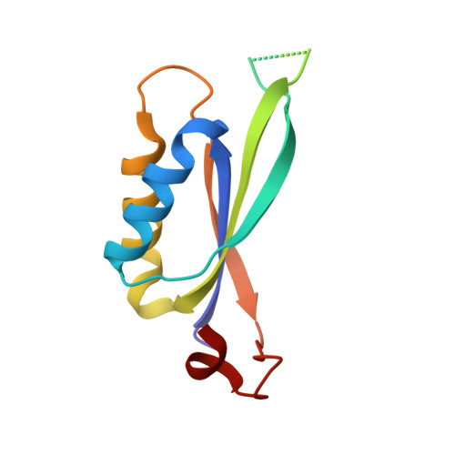
    PII constitutes a family of signal transduction proteins that act as nitrogen sensors in microorganisms and plants. Mycobacterium tuberculosis (Mtb) has a single homologue of PII whose precise role has as yet not been explored. We have solved the crystal structures of the Mtb PII protein in its apo and ATP bound forms to 1.4 and 2.4 A resolutions, respectively. The protein forms a trimeric assembly in the crystal lattice and folds similarly to the other PII family proteins. The Mtb PII:ATP binary complex structure reveals three ATP molecules per trimer, each bound between the base of the T-loop of one subunit and the C-loop of the neighboring subunit. In contrast to the apo structure, at least one subunit of the binary complex structure contains a completely ordered T-loop indicating that ATP binding plays a role in orienting this loop region towards target proteins like the ammonium transporter, AmtB. Arg38 of the T-loop makes direct contact with the gamma-phosphate of the ATP molecule replacing the Mg(2+) position seen in the Methanococcus jannaschii GlnK1 structure. The C-loop of a neighboring subunit encloses the other side of the ATP molecule, placing the GlnK specific C-terminal 3(10) helix in the vicinity. Homology modeling studies with the E. coli GlnK:AmtB complex reveal that Mtb PII could form a complex similar to the complex in E. coli. The structural conservation and operon organization suggests that the Mtb PII gene encodes for a GlnK protein and might play a key role in the nitrogen regulatory pathway.


  • Organizational Affiliation

    Department of Chemistry, Texas A&M University, College Station, Texas 77843-2128, USA.


Macromolecules
Find similar proteins by:  (by identity cutoff)  |  3D Structure
Entity ID: 1
MoleculeChains Sequence LengthOrganismDetailsImage
Nitrogen regulatory protein P-II
A, B, C
114Mycobacterium tuberculosisMutation(s): 0 
Gene Names: glnBMT2987MTCY338.08cRv2919c
UniProt
Find proteins for P9WN31 (Mycobacterium tuberculosis (strain ATCC 25618 / H37Rv))
Explore P9WN31 
Go to UniProtKB:  P9WN31
Entity Groups  
Sequence Clusters30% Identity50% Identity70% Identity90% Identity95% Identity100% Identity
UniProt GroupP9WN31
Sequence Annotations
Expand
  • Reference Sequence
Small Molecules
Binding Affinity Annotations 
IDSourceBinding Affinity
ATP Binding MOAD:  3LF0 Kd: 1930 (nM) from 1 assay(s)
PDBBind:  3LF0 Kd: 1930 (nM) from 1 assay(s)
Experimental Data & Validation

Experimental Data

  • Method: X-RAY DIFFRACTION
  • Resolution: 2.40 Å
  • R-Value Free: 0.272 
  • R-Value Work: 0.204 
  • R-Value Observed: 0.208 
  • Space Group: P 43 21 2
Unit Cell:
Length ( Å )Angle ( ˚ )
a = 69.742α = 90
b = 69.742β = 90
c = 146.682γ = 90
Software Package:
Software NamePurpose
HKL-2000data collection
CCP4model building
REFMACrefinement
HKL-2000data reduction
HKL-2000data scaling
CCP4phasing

Structure Validation

View Full Validation Report


Ligand Structure Quality Assessment 


Entry History 

Deposition Data

Revision History  (Full details and data files)

  • Version 1.0: 2010-07-07
    Type: Initial release
  • Version 1.1: 2011-07-13
    Changes: Version format compliance
  • Version 1.2: 2023-09-06
    Changes: Data collection, Database references, Derived calculations, Refinement description