3LCU

Crystal Structure of Antibiotic related Methyltransferase


Experimental Data Snapshot

  • Method: X-RAY DIFFRACTION
  • Resolution: 2.10 Å
  • R-Value Free: 0.259 
  • R-Value Work: 0.205 
  • R-Value Observed: 0.205 

wwPDB Validation   3D Report Full Report


Ligand Structure Quality Assessment 


This is version 1.2 of the entry. See complete history


Literature

Structural basis for the methylation of G1405 in 16S rRNA by aminoglycoside resistance methyltransferase Sgm from an antibiotic producer: a diversity of active sites in m7G methyltransferases.

Husain, N.Tkaczuk, K.L.Tulsidas, S.R.Kaminska, K.H.Cubrilo, S.Maravic-Vlahovicek, G.Bujnicki, J.M.Sivaraman, J.

(2010) Nucleic Acids Res 

  • DOI: https://doi.org/10.1093/nar/gkq122
  • Primary Citation of Related Structures:  
    3LCU, 3LCV

  • PubMed Abstract: 

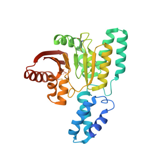
    Sgm (Sisomicin-gentamicin methyltransferase) from antibiotic-producing bacterium Micromonospora zionensis is an enzyme that confers resistance to aminoglycosides like gentamicin and sisomicin by specifically methylating G1405 in bacterial 16S rRNA. Sgm belongs to the aminoglycoside resistance methyltransferase (Arm) family of enzymes that have been recently found to spread by horizontal gene transfer among disease-causing bacteria. Structural characterization of Arm enzymes is the key to understand their mechanism of action and to develop inhibitors that would block their activity. Here we report the structure of Sgm in complex with cofactors S-adenosylmethionine (AdoMet) and S-adenosylhomocysteine (AdoHcy) at 2.0 and 2.1 A resolution, respectively, and results of mutagenesis and rRNA footprinting, and protein-substrate docking. We propose the mechanism of methylation of G1405 by Sgm and compare it with other m(7)G methyltransferases, revealing a surprising diversity of active sites and binding modes for the same basic reaction of RNA modification. This analysis can serve as a stepping stone towards developing drugs that would specifically block the activity of Arm methyltransferases and thereby re-sensitize pathogenic bacteria to aminoglycoside antibiotics.


  • Organizational Affiliation

    Department of Biological Sciences, 14 Science drive 4, National University of Singapore, Singapore.


Macromolecules
Find similar proteins by:  (by identity cutoff)  |  3D Structure
Entity ID: 1
MoleculeChains Sequence LengthOrganismDetailsImage
Sisomicin-gentamicin resistance methylase Sgm281Micromonospora zionensisMutation(s): 0 
UniProt
Find proteins for Q7M0R2 (Micromonospora zionensis)
Explore Q7M0R2 
Go to UniProtKB:  Q7M0R2
Entity Groups  
Sequence Clusters30% Identity50% Identity70% Identity90% Identity95% Identity100% Identity
UniProt GroupQ7M0R2
Sequence Annotations
Expand
  • Reference Sequence
Small Molecules
Ligands 1 Unique
IDChains Name / Formula / InChI Key2D Diagram3D Interactions
SAH
Query on SAH

Download Ideal Coordinates CCD File 
B [auth A]S-ADENOSYL-L-HOMOCYSTEINE
C14 H20 N6 O5 S
ZJUKTBDSGOFHSH-WFMPWKQPSA-N
Modified Residues  1 Unique
IDChains TypeFormula2D DiagramParent
MSE
Query on MSE
A
L-PEPTIDE LINKINGC5 H11 N O2 SeMET
Binding Affinity Annotations 
IDSourceBinding Affinity
SAH PDBBind:  3LCU Kd: 3.00e+5 (nM) from 1 assay(s)
Binding MOAD:  3LCU Kd: 3.00e+5 (nM) from 1 assay(s)
Experimental Data & Validation

Experimental Data

  • Method: X-RAY DIFFRACTION
  • Resolution: 2.10 Å
  • R-Value Free: 0.259 
  • R-Value Work: 0.205 
  • R-Value Observed: 0.205 
  • Space Group: P 1 21 1
Unit Cell:
Length ( Å )Angle ( ˚ )
a = 36.087α = 90
b = 68.891β = 93.47
c = 52.201γ = 90
Software Package:
Software NamePurpose
DENZOdata reduction
SCALEPACKdata scaling
CNSrefinement
PDB_EXTRACTdata extraction

Structure Validation

View Full Validation Report


Ligand Structure Quality Assessment 


Entry History 

Deposition Data

Revision History  (Full details and data files)

  • Version 1.0: 2010-06-02
    Type: Initial release
  • Version 1.1: 2011-07-13
    Changes: Version format compliance
  • Version 1.2: 2017-11-01
    Changes: Refinement description