3LC0

Histidyl-tRNA synthetase from Trypanosoma cruzi (Histidine complex)


Experimental Data Snapshot

  • Method: X-RAY DIFFRACTION
  • Resolution: 1.80 Å
  • R-Value Free: 0.211 
  • R-Value Work: 0.193 

wwPDB Validation   3D Report Full Report


Ligand Structure Quality Assessment 


This is version 1.3 of the entry. See complete history


Literature

Crystal Structures of Trypanosomal Histidyl-tRNA Synthetase Illuminate Differences between Eukaryotic and Prokaryotic Homologs.

Merritt, E.A.Arakaki, T.L.Gillespie, J.R.Larson, E.T.Kelley, A.Mueller, N.Napuli, A.J.Kim, J.Zhang, L.Verlinde, C.L.Fan, E.Zucker, F.Buckner, F.S.Van Voorhis, W.C.Hol, W.G.

(2010) J Mol Biol 397: 481-494

  • DOI: https://doi.org/10.1016/j.jmb.2010.01.051
  • Primary Citation of Related Structures:  
    3HRI, 3HRK, 3LC0

  • PubMed Abstract: 

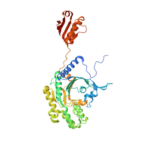
    Crystal structures of histidyl-tRNA synthetase (HisRS) from the eukaryotic parasites Trypanosoma brucei and Trypanosoma cruzi provide a first structural view of a eukaryotic form of this enzyme and reveal differences from bacterial homologs. HisRSs in general contain an extra domain inserted between conserved motifs 2 and 3 of the Class II aminoacyl-tRNA synthetase catalytic core. The current structures show that the three-dimensional topology of this domain is very different in bacterial and archaeal/eukaryotic forms of the enzyme. Comparison of apo and histidine-bound trypanosomal structures indicates substantial active-site rearrangement upon histidine binding but relatively little subsequent rearrangement after reaction of histidine with ATP to form the enzyme's first reaction product, histidyladenylate. The specific residues involved in forming the binding pocket for the adenine moiety differ substantially both from the previously characterized binding site in bacterial structures and from the homologous residues in human HisRSs. The essentiality of the single HisRS gene in T. brucei is shown by a severe depression of parasite growth rate that results from even partial suppression of expression by RNA interference.


  • Organizational Affiliation

    Department of Biochemistry, University of Washington, Seattle, WA 98195, USA. merritt@u.washington.edu


Macromolecules
Find similar proteins by:  (by identity cutoff)  |  3D Structure
Entity ID: 1
MoleculeChains Sequence LengthOrganismDetailsImage
Histidyl-tRNA synthetase456Trypanosoma cruziMutation(s): 0 
Gene Names: Tc00.1047053507019.40
EC: 6.1.1.21
UniProt
Find proteins for Q4DA54 (Trypanosoma cruzi (strain CL Brener))
Explore Q4DA54 
Go to UniProtKB:  Q4DA54
Entity Groups  
Sequence Clusters30% Identity50% Identity70% Identity90% Identity95% Identity100% Identity
UniProt GroupQ4DA54
Sequence Annotations
Expand
  • Reference Sequence
Small Molecules
Ligands 1 Unique
IDChains Name / Formula / InChI Key2D Diagram3D Interactions
HIS
Query on HIS

Download Ideal Coordinates CCD File 
B [auth A]HISTIDINE
C6 H10 N3 O2
HNDVDQJCIGZPNO-YFKPBYRVSA-O
Experimental Data & Validation

Experimental Data

  • Method: X-RAY DIFFRACTION
  • Resolution: 1.80 Å
  • R-Value Free: 0.211 
  • R-Value Work: 0.193 
  • Space Group: C 1 2 1
Unit Cell:
Length ( Å )Angle ( ˚ )
a = 90.03α = 90
b = 118.84β = 132.57
c = 65.98γ = 90
Software Package:
Software NamePurpose
SCALAdata scaling
REFMACrefinement
PDB_EXTRACTdata extraction
MOSFLMdata reduction

Structure Validation

View Full Validation Report


Ligand Structure Quality Assessment 


Entry History 

Deposition Data

Revision History  (Full details and data files)

  • Version 1.0: 2010-01-19
    Type: Initial release
  • Version 1.1: 2011-07-13
    Changes: Advisory, Version format compliance
  • Version 1.2: 2017-11-01
    Changes: Refinement description
  • Version 1.3: 2024-02-21
    Changes: Data collection, Database references, Derived calculations