


The Crystal Structure of smu.665 from Streptococcus mutans UA159
Su, X.-D., Liu, X., Wu, C.W.To be published.
Experimental Data Snapshot
Entity ID: 1 | |||||
|---|---|---|---|---|---|
| Molecule | Chains | Sequence Length | Organism | Details | Image |
| Acetylglutamate kinase | 279 | Streptococcus mutans UA159 | Mutation(s): 0 Gene Names: smu.665 EC: 2.7.2.8 | 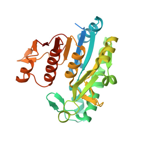 | |
UniProt | |||||
Find proteins for Q8DV44 (Streptococcus mutans serotype c (strain ATCC 700610 / UA159)) Explore Q8DV44 Go to UniProtKB: Q8DV44 | |||||
Entity Groups | |||||
| Sequence Clusters | 30% Identity50% Identity70% Identity90% Identity95% Identity100% Identity | ||||
| UniProt Group | Q8DV44 | ||||
Sequence AnnotationsExpand | |||||
| |||||
| Ligands 3 Unique | |||||
|---|---|---|---|---|---|
| ID | Chains | Name / Formula / InChI Key | 2D Diagram | 3D Interactions | |
| ADP Query on ADP | B [auth A] | ADENOSINE-5'-DIPHOSPHATE C10 H15 N5 O10 P2 XTWYTFMLZFPYCI-KQYNXXCUSA-N |  | ||
| NLG Query on NLG | C [auth A] | N-ACETYL-L-GLUTAMATE C7 H11 N O5 RFMMMVDNIPUKGG-YFKPBYRVSA-N |  | ||
| MG Query on MG | D [auth A] | MAGNESIUM ION Mg JLVVSXFLKOJNIY-UHFFFAOYSA-N |  | ||
| Length ( Å ) | Angle ( ˚ ) |
|---|---|
| a = 57.19 | α = 90 |
| b = 94.76 | β = 90 |
| c = 47.58 | γ = 90 |
| Software Name | Purpose |
|---|---|
| MAR345dtb | data collection |
| MOLREP | phasing |
| REFMAC | refinement |
| XDS | data reduction |
| XDS | data scaling |
RCSB PDB (citation) is hosted by
RCSB PDB is a member of the

