


A dimeric mutant of DHDPS from Mycobacterium tuberculosis
Evans, G.L., Schuldt, L., Griffin, M.D., Perugini, M.A., Jamerson, G.B., Devenish, S.R., Weiss, M.S., Gerrard, J.A.To be published.
Experimental Data Snapshot
wwPDB Validation 3D Report Full Report

Entity ID: 1 | |||||
|---|---|---|---|---|---|
| Molecule | Chains | Sequence Length | Organism | Details | Image |
| Dihydrodipicolinate synthase | 304 | Mycobacterium tuberculosis | Mutation(s): 1 Gene Names: dapA, MT2823, MTV002.18c, Rv2753c EC: 4.2.1.52 | 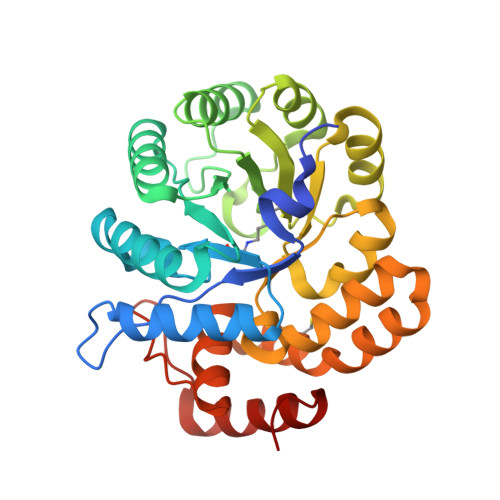 | |
UniProt | |||||
Find proteins for P9WP25 (Mycobacterium tuberculosis (strain ATCC 25618 / H37Rv)) Explore P9WP25 Go to UniProtKB: P9WP25 | |||||
Entity Groups | |||||
| Sequence Clusters | 30% Identity50% Identity70% Identity90% Identity95% Identity100% Identity | ||||
| UniProt Group | P9WP25 | ||||
Sequence AnnotationsExpand | |||||
| |||||
| Ligands 6 Unique | |||||
|---|---|---|---|---|---|
| ID | Chains | Name / Formula / InChI Key | 2D Diagram | 3D Interactions | |
| SO4 Query on SO4 | FA [auth C] FB [auth F] G [auth A] GA [auth C] GB [auth F] | SULFATE ION O4 S QAOWNCQODCNURD-UHFFFAOYSA-L |  | ||
| GOL Query on GOL | CA [auth B] CB [auth E] DB [auth E] EA [auth B] IA [auth C] | GLYCEROL C3 H8 O3 PEDCQBHIVMGVHV-UHFFFAOYSA-N |  | ||
| PYR Query on PYR | DA [auth B] | PYRUVIC ACID C3 H4 O3 LCTONWCANYUPML-UHFFFAOYSA-N |  | ||
| BME Query on BME | Q [auth A] | BETA-MERCAPTOETHANOL C2 H6 O S DGVVWUTYPXICAM-UHFFFAOYSA-N |  | ||
| ACT Query on ACT | AA [auth B] AB [auth E] BA [auth B] BB [auth E] HB [auth F] | ACETATE ION C2 H3 O2 QTBSBXVTEAMEQO-UHFFFAOYSA-M |  | ||
| CL Query on CL | EB [auth E], UA [auth D], VA [auth D] | CHLORIDE ION Cl VEXZGXHMUGYJMC-UHFFFAOYSA-M |  | ||
| Modified Residues 2 Unique | |||||
|---|---|---|---|---|---|
| ID | Chains | Type | Formula | 2D Diagram | Parent |
| CME Query on CME | A, B, C, D, E A, B, C, D, E, F | L-PEPTIDE LINKING | C5 H11 N O3 S2 |  | CYS |
| KPI Query on KPI | A, B, C, D, E A, B, C, D, E, F | L-PEPTIDE LINKING | C9 H16 N2 O4 |  | LYS |
| Length ( Å ) | Angle ( ˚ ) |
|---|---|
| a = 188.83 | α = 90 |
| b = 188.83 | β = 90 |
| c = 130.43 | γ = 90 |
| Software Name | Purpose |
|---|---|
| MxCuBE | data collection |
| MOLREP | phasing |
| REFMAC | refinement |
| DENZO | data reduction |
| SCALEPACK | data scaling |
RCSB PDB (citation) is hosted by
RCSB PDB is a member of the

