3L1P

POU protein:DNA complex


Experimental Data Snapshot

  • Method: X-RAY DIFFRACTION
  • Resolution: 2.80 Å
  • R-Value Free: 0.257 
  • R-Value Work: 0.224 
  • R-Value Observed: 0.226 

wwPDB Validation   3D Report Full Report


This is version 1.4 of the entry. See complete history


Literature

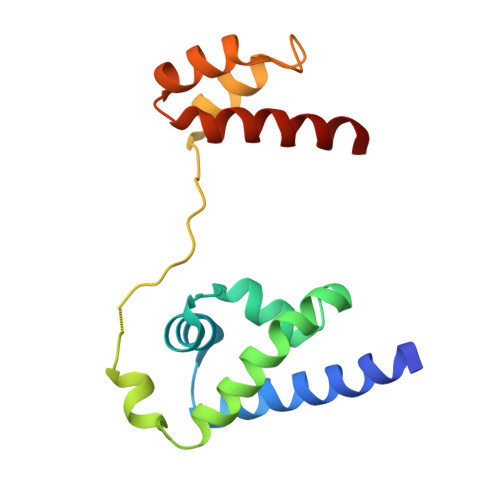
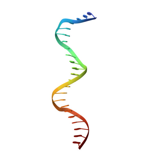
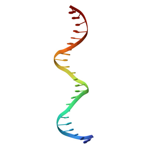
A unique Oct4 interface is crucial for reprogramming to pluripotency

Esch, D.Vahokoski, J.Groves, M.R.Pogenberg, V.Cojocaru, V.Vom Bruch, H.Han, D.Drexler, H.C.A.Arauzo-Bravo, M.J.Ng, C.K.L.Jauch, R.Wilmanns, M.Scholer, H.R.

(2013) Nat Cell Biol 15: 295-301

  • DOI: https://doi.org/10.1038/ncb2680
  • Primary Citation of Related Structures:  
    3L1P

  • PubMed Abstract: 

    Terminally differentiated cells can be reprogrammed to pluripotency by the forced expression of Oct4, Sox2, Klf4 and c-Myc. However, it remains unknown how this leads to the multitude of epigenetic changes observed during the reprogramming process. Interestingly, Oct4 is the only factor that cannot be replaced by other members of the same family to induce pluripotency. To understand the unique role of Oct4 in reprogramming, we determined the structure of its POU domain bound to DNA. We show that the linker between the two DNA-binding domains is structured as an α-helix and exposed to the protein's surface, in contrast to the unstructured linker of Oct1. Point mutations in this α-helix alter or abolish the reprogramming activity of Oct4, but do not affect its other fundamental properties. On the basis of mass spectrometry studies of the interactome of wild-type and mutant Oct4, we propose that the linker functions as a protein-protein interaction interface and plays a crucial role during reprogramming by recruiting key epigenetic players to Oct4 target genes. Thus, we provide molecular insights to explain how Oct4 contributes to the reprogramming process.


  • Organizational Affiliation

    Department for Cell and Developmental Biology, Max Planck Institute for Molecular Biomedicine, Röntgenstrasse 20, Münster D-48149, Germany.


Macromolecules

Find similar proteins by:  (by identity cutoff)  |  3D Structure
Entity ID: 1
MoleculeChains Sequence LengthOrganismDetailsImage
POU domain, class 5, transcription factor 1
A, B
155Mus musculusMutation(s): 5 
Gene Names: PO5F1
UniProt
Find proteins for P20263 (Mus musculus)
Explore P20263 
Go to UniProtKB:  P20263
Entity Groups  
Sequence Clusters30% Identity50% Identity70% Identity90% Identity95% Identity100% Identity
UniProt GroupP20263
Sequence Annotations
Expand
  • Reference Sequence

Find similar nucleic acids by:  Sequence   |   3D Structure  

Entity ID: 2
MoleculeChains LengthOrganismImage
DNA (5'-D(*TP*CP*CP*AP*CP*AP*TP*TP*TP*GP*AP*AP*AP*GP*GP*CP*AP*AP*AP*TP*GP*GP*A)-3')C [auth N]23N/A
Sequence Annotations
Expand
  • Reference Sequence

Find similar nucleic acids by:  Sequence   |   3D Structure  

Entity ID: 3
MoleculeChains LengthOrganismImage
DNA (5'-D(*AP*TP*CP*CP*AP*TP*TP*TP*GP*CP*CP*TP*TP*TP*CP*AP*AP*AP*TP*GP*TP*GP*G)-3')D [auth M]23N/A
Sequence Annotations
Expand
  • Reference Sequence
Experimental Data & Validation

Experimental Data

  • Method: X-RAY DIFFRACTION
  • Resolution: 2.80 Å
  • R-Value Free: 0.257 
  • R-Value Work: 0.224 
  • R-Value Observed: 0.226 
  • Space Group: P 41
Unit Cell:
Length ( Å )Angle ( ˚ )
a = 49.25α = 90
b = 49.25β = 90
c = 265.48γ = 90
Software Package:
Software NamePurpose
DNAdata collection
PHASERphasing
REFMACrefinement
XDSdata reduction
XSCALEdata scaling

Structure Validation

View Full Validation Report


Entry History 

Deposition Data

Revision History  (Full details and data files)

  • Version 1.0: 2010-12-15
    Type: Initial release
  • Version 1.1: 2011-07-13
    Changes: Version format compliance
  • Version 1.2: 2013-06-05
    Changes: Database references
  • Version 1.3: 2018-02-07
    Changes: Experimental preparation
  • Version 1.4: 2023-11-01
    Changes: Data collection, Database references, Refinement description