3KXZ

The complex crystal structure of LCK with a probe molecule w259


Experimental Data Snapshot

  • Method: X-RAY DIFFRACTION
  • Resolution: 2.37 Å
  • R-Value Free: 0.261 
  • R-Value Work: 0.192 

wwPDB Validation   3D Report Full Report


Ligand Structure Quality Assessment 


This is version 1.1 of the entry. See complete history


Literature

Novel synthesis and structural characterization of a high-affinity paramagnetic kinase probe for the identification of non-ATP site binders by nuclear magnetic resonance.

Moy, F.J.Lee, A.Gavrin, L.K.Xu, Z.B.Sievers, A.Kieras, E.Stochaj, W.Mosyak, L.McKew, J.Tsao, D.H.

(2010) J Med Chem 53: 1238-1249

  • DOI: https://doi.org/10.1021/jm901525b
  • Primary Citation of Related Structures:  
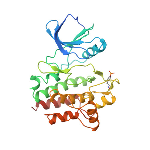
    3KXZ

  • PubMed Abstract: 

    To aid in the pursuit of selective kinase inhibitors, we have developed a unique ATP site binder tool for the detection of binders outside the ATP site by nuclear magnetic resonance (NMR). We report here the novel synthesis that led to this paramagnetic spin-labeled pyrazolopyrimidine probe (1), which exhibits nanomolar inhibitory activity against multiple kinases. We demonstrate the application of this probe by performing NMR binding experiments with Lck and Src kinases and utilize it to detect the binding of two compounds proximal to the ATP site. The complex structure of the probe with Lck is also presented, revealing how the probe fits in the ATP site and the specific interactions it has with the protein. We believe that this spin-labeled probe is a valuable tool that holds broad applicability in a screen for non-ATP site binders.


  • Organizational Affiliation

    Structural Biology and Computational Chemistry, Wyeth Research, 200 CambridgePark Drive, Cambridge, Massachusetts 02140, USA.


Macromolecules
Find similar proteins by:  (by identity cutoff)  |  3D Structure
Entity ID: 1
MoleculeChains Sequence LengthOrganismDetailsImage
Proto-oncogene tyrosine-protein kinase LCK287Homo sapiensMutation(s): 0 
Gene Names: LCK
EC: 2.7.10.2
UniProt & NIH Common Fund Data Resources
Find proteins for P06239 (Homo sapiens)
Explore P06239 
Go to UniProtKB:  P06239
PHAROS:  P06239
GTEx:  ENSG00000182866 
Entity Groups  
Sequence Clusters30% Identity50% Identity70% Identity90% Identity95% Identity100% Identity
UniProt GroupP06239
Sequence Annotations
Expand
  • Reference Sequence
Small Molecules
Ligands 3 Unique
IDChains Name / Formula / InChI Key2D Diagram3D Interactions
925
Query on 925

Download Ideal Coordinates CCD File 
D [auth A]3-[7-[(3-hydroxyphenyl)amino]pyrazolo[1,5-a]pyrimidin-2-yl]-N-(1-hydroxy-2,2,6,6-tetramethyl-piperidin-4-yl)benzamide
C28 H32 N6 O3
WZEJOLDIQBJKQR-UHFFFAOYSA-N
SO4
Query on SO4

Download Ideal Coordinates CCD File 
B [auth A]SULFATE ION
O4 S
QAOWNCQODCNURD-UHFFFAOYSA-L
CA
Query on CA

Download Ideal Coordinates CCD File 
C [auth A]CALCIUM ION
Ca
BHPQYMZQTOCNFJ-UHFFFAOYSA-N
Modified Residues  1 Unique
IDChains TypeFormula2D DiagramParent
PTR
Query on PTR
A
L-PEPTIDE LINKINGC9 H12 N O6 PTYR
Binding Affinity Annotations 
IDSourceBinding Affinity
925 Binding MOAD:  3KXZ IC50: 35 (nM) from 1 assay(s)
PDBBind:  3KXZ IC50: 35 (nM) from 1 assay(s)
PTR BindingDB:  3KXZ Kd: 4.00e+5 (nM) from 1 assay(s)
Experimental Data & Validation

Experimental Data

  • Method: X-RAY DIFFRACTION
  • Resolution: 2.37 Å
  • R-Value Free: 0.261 
  • R-Value Work: 0.192 
  • Space Group: P 21 21 21
Unit Cell:
Length ( Å )Angle ( ˚ )
a = 42.012α = 90
b = 73.784β = 90
c = 84.717γ = 90
Software Package:
Software NamePurpose
ADSCdata collection
PHASERphasing
PHENIXrefinement
HKL-2000data reduction
SCALEPACKdata scaling

Structure Validation

View Full Validation Report


Ligand Structure Quality Assessment 


Entry History 

Deposition Data

Revision History  (Full details and data files)

  • Version 1.0: 2010-06-23
    Type: Initial release
  • Version 1.1: 2011-07-13
    Changes: Version format compliance