3KX0

Crystal Structure of the PAS domain of Rv1364c


Experimental Data Snapshot

  • Method: X-RAY DIFFRACTION
  • Resolution: 2.30 Å
  • R-Value Free: 0.274 
  • R-Value Work: 0.233 
  • R-Value Observed: 0.235 

wwPDB Validation   3D Report Full Report


This is version 1.1 of the entry. See complete history


Literature

Role of a PAS sensor domain in the Mycobacterium tuberculosis transcription regulator Rv1364c

Jaiswal, R.K.Manjeera, G.Gopal, B.

(2010) Biochem Biophys Res Commun 398: 342-349

  • DOI: https://doi.org/10.1016/j.bbrc.2010.06.027
  • Primary Citation of Related Structures:  
    3KX0

  • PubMed Abstract: 

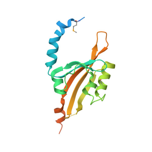
    The Mycobacterium tuberculosis transcriptional regulator Rv1364c regulates the activity of the stress response sigma factor sigma(F). This multi-domain protein has several components: a signaling PAS domain and an effector segment comprising of a phosphatase, a kinase and an anti-anti-sigma factor domain. Based on Small Angle X-ray Scattering (SAXS) data, Rv1364c was recently shown to be a homo-dimer and adopt an elongated conformation in solution. The PAS domain could not be modeled into the structural envelope due to poor sequence similarity with known PAS proteins. The crystal structure of the PAS domain described here provides a structural basis for the dimerization of Rv1364c. It thus appears likely that the PAS domain regulates the anti-sigma activity of Rv1364c by oligomerization. A structural comparison with other characterized PAS domains reveal several sequence and conformational features that could facilitate ligand binding - a feature which suggests that the function of Rv1364c could potentially be governed by specific cellular signals or metabolic cues.


  • Organizational Affiliation

    Molecular Biophysics Unit, Indian Institute of Science, Bangalore 560012, India.


Macromolecules
Find similar proteins by:  (by identity cutoff)  |  3D Structure
Entity ID: 1
MoleculeChains Sequence LengthOrganismDetailsImage
Uncharacterized protein Rv1364c/MT1410A [auth X]185Mycobacterium tuberculosisMutation(s): 0 
Gene Names: Rv1364c
UniProt
Find proteins for P9WLZ7 (Mycobacterium tuberculosis (strain ATCC 25618 / H37Rv))
Explore P9WLZ7 
Go to UniProtKB:  P9WLZ7
Entity Groups  
Sequence Clusters30% Identity50% Identity70% Identity90% Identity95% Identity100% Identity
UniProt GroupP9WLZ7
Sequence Annotations
Expand
  • Reference Sequence
Small Molecules
Ligands 1 Unique
IDChains Name / Formula / InChI Key2D Diagram3D Interactions
IPA
Query on IPA

Download Ideal Coordinates CCD File 
B [auth X]ISOPROPYL ALCOHOL
C3 H8 O
KFZMGEQAYNKOFK-UHFFFAOYSA-N
Modified Residues  1 Unique
IDChains TypeFormula2D DiagramParent
MSE
Query on MSE
A [auth X]L-PEPTIDE LINKINGC5 H11 N O2 SeMET
Experimental Data & Validation

Experimental Data

  • Method: X-RAY DIFFRACTION
  • Resolution: 2.30 Å
  • R-Value Free: 0.274 
  • R-Value Work: 0.233 
  • R-Value Observed: 0.235 
  • Space Group: P 62 2 2
Unit Cell:
Length ( Å )Angle ( ˚ )
a = 59.609α = 90
b = 59.609β = 90
c = 173.327γ = 120
Software Package:
Software NamePurpose
SOLVEphasing
PHENIXrefinement
MOSFLMdata reduction
SCALAdata scaling

Structure Validation

View Full Validation Report


Entry History 

Deposition Data

Revision History  (Full details and data files)

  • Version 1.0: 2010-06-23
    Type: Initial release
  • Version 1.1: 2011-07-13
    Changes: Version format compliance