3KV2

HIGH RESOLUTION STRUCTURE OF HUMAN ARGINASE I IN COMPLEX WITH THE STRONG INHIBITOR N(omega)-hydroxy-nor-L-arginine (nor-NOHA)


Experimental Data Snapshot

  • Method: X-RAY DIFFRACTION
  • Resolution: 1.55 Å
  • R-Value Free: 0.178 
  • R-Value Work: 0.144 
  • R-Value Observed: 0.144 

wwPDB Validation   3D Report Full Report


Ligand Structure Quality Assessment 


This is version 1.2 of the entry. See complete history


Literature

Inhibition of human arginase I by substrate and product analogues.

Di Costanzo, L.Ilies, M.Thorn, K.J.Christianson, D.W.

(2010) Arch Biochem Biophys 496: 101-108

  • DOI: https://doi.org/10.1016/j.abb.2010.02.004
  • Primary Citation of Related Structures:  
    3KV2, 3LP4, 3LP7

  • PubMed Abstract: 

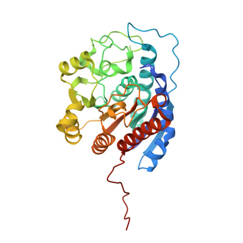
    Human arginase I is a binuclear manganese metalloenzyme that catalyzes the hydrolysis of L-arginine to generate L-ornithine and urea. We demonstrate that N-hydroxy-L-arginine (NOHA) binds to this enzyme with K(d)=3.6 microM, and nor-N-hydroxy-L-arginine (nor-NOHA) binds with K(d)=517 nM (surface plasmon resonance) or K(d) approximately 50 nM (isothermal titration calorimetry). Crystals of human arginase I complexed with NOHA and nor-NOHA afford 2.04 and 1.55 A resolution structures, respectively, which are significantly improved in comparison with previously-determined structures of the corresponding complexes with rat arginase I. Higher resolution structures clarify the binding interactions of the inhibitors. Finally, the crystal structure of the complex with L-lysine (K(d)=13 microM) is reported at 1.90 A resolution. This structure confirms the importance of hydrogen bond interactions with inhibitor alpha-carboxylate and alpha-amino groups as key specificity determinants of amino acid recognition in the arginase active site.


  • Organizational Affiliation

    Roy and Diana Vagelos Laboratories, Department of Chemistry, University of Pennsylvania, Philadelphia, PA 19104-6323, USA.


Macromolecules
Find similar proteins by:  (by identity cutoff)  |  3D Structure
Entity ID: 1
MoleculeChains Sequence LengthOrganismDetailsImage
Arginase-1
A, B
322Homo sapiensMutation(s): 0 
Gene Names: ARG1
EC: 3.5.3.1
UniProt & NIH Common Fund Data Resources
Find proteins for P05089 (Homo sapiens)
Explore P05089 
Go to UniProtKB:  P05089
PHAROS:  P05089
GTEx:  ENSG00000118520 
Entity Groups  
Sequence Clusters30% Identity50% Identity70% Identity90% Identity95% Identity100% Identity
UniProt GroupP05089
Sequence Annotations
Expand
  • Reference Sequence
Small Molecules
Binding Affinity Annotations 
IDSourceBinding Affinity
NNH PDBBind:  3KV2 Kd: 47.51 (nM) from 1 assay(s)
BindingDB:  3KV2 Kd: min: 47, max: 517 (nM) from 2 assay(s)
IC50: min: 800, max: 6000 (nM) from 5 assay(s)
Binding MOAD:  3KV2 Kd: 47.51 (nM) from 1 assay(s)
Experimental Data & Validation

Experimental Data

  • Method: X-RAY DIFFRACTION
  • Resolution: 1.55 Å
  • R-Value Free: 0.178 
  • R-Value Work: 0.144 
  • R-Value Observed: 0.144 
  • Space Group: P 3
Unit Cell:
Length ( Å )Angle ( ˚ )
a = 90.497α = 90
b = 90.497β = 90
c = 69.604γ = 120
Software Package:
Software NamePurpose
CNSrefinement
HKL-2000data reduction
SCALEPACKdata scaling

Structure Validation

View Full Validation Report


Ligand Structure Quality Assessment 


Entry History 

Deposition Data

Revision History  (Full details and data files)

  • Version 1.0: 2009-12-22
    Type: Initial release
  • Version 1.1: 2011-07-13
    Changes: Version format compliance
  • Version 1.2: 2023-09-06
    Changes: Data collection, Database references, Derived calculations, Refinement description