3KR8

Human tankyrase 2 - catalytic PARP domain in complex with an inhibitor XAV939


Experimental Data Snapshot

  • Method: X-RAY DIFFRACTION
  • Resolution: 2.10 Å
  • R-Value Free: 0.255 
  • R-Value Work: 0.198 
  • R-Value Observed: 0.201 

wwPDB Validation   3D Report Full Report


Ligand Structure Quality Assessment 


This is version 1.2 of the entry. See complete history


Literature

Structural basis for the interaction between tankyrase-2 and a potent Wnt-signaling inhibitor.

Karlberg, T.Markova, N.Johansson, I.Hammarstrom, M.Schutz, P.Weigelt, J.Schuler, H.

(2010) J Med Chem 53: 5352-5355

  • DOI: https://doi.org/10.1021/jm100249w
  • Primary Citation of Related Structures:  
    3KR7, 3KR8

  • PubMed Abstract: 

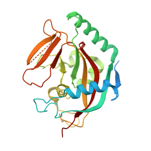
    We report two crystal structures of the PARP domain of human tankyrase-2 (TNKS2). Tankyrases are involved in fundamental cellular processes such as telomere homeostasis and Wnt signaling. The complex of TNKS2 with the potent inhibitor XAV939 provides insights into the molecular basis of the strong interaction and suggests routes for further development of tankyrase inhibitors.


  • Organizational Affiliation

    Structural Genomics Consortium, Department of Medical Biochemistry and Biophysics, Karolinska Institutet, S-17177 Stockholm, Sweden.


Macromolecules
Find similar proteins by:  (by identity cutoff)  |  3D Structure
Entity ID: 1
MoleculeChains Sequence LengthOrganismDetailsImage
Tankyrase-2A,
B [auth C]
240Homo sapiensMutation(s): 0 
Gene Names: PARP5BTANK2TNKLTNKS2
EC: 2.4.2.30
UniProt & NIH Common Fund Data Resources
Find proteins for Q9H2K2 (Homo sapiens)
Explore Q9H2K2 
Go to UniProtKB:  Q9H2K2
PHAROS:  Q9H2K2
GTEx:  ENSG00000107854 
Entity Groups  
Sequence Clusters30% Identity50% Identity70% Identity90% Identity95% Identity100% Identity
UniProt GroupQ9H2K2
Sequence Annotations
Expand
  • Reference Sequence
Small Molecules
Ligands 4 Unique
IDChains Name / Formula / InChI Key2D Diagram3D Interactions
XAV
Query on XAV

Download Ideal Coordinates CCD File 
D [auth A],
I [auth C]
2-[4-(trifluoromethyl)phenyl]-7,8-dihydro-5H-thiopyrano[4,3-d]pyrimidin-4-ol
C14 H11 F3 N2 O S
KLGQSVMIPOVQAX-UHFFFAOYSA-N
SO4
Query on SO4

Download Ideal Coordinates CCD File 
E [auth A],
F [auth A],
J [auth C],
K [auth C]
SULFATE ION
O4 S
QAOWNCQODCNURD-UHFFFAOYSA-L
GOL
Query on GOL

Download Ideal Coordinates CCD File 
G [auth A],
L [auth C]
GLYCEROL
C3 H8 O3
PEDCQBHIVMGVHV-UHFFFAOYSA-N
ZN
Query on ZN

Download Ideal Coordinates CCD File 
C [auth A],
H [auth C]
ZINC ION
Zn
PTFCDOFLOPIGGS-UHFFFAOYSA-N
Binding Affinity Annotations 
IDSourceBinding Affinity
XAV BindingDB:  3KR8 Kd: min: 4.2, max: 14 (nM) from 3 assay(s)
IC50: min: 2.1, max: 17 (nM) from 9 assay(s)
Binding MOAD:  3KR8 Kd: 8 (nM) from 1 assay(s)
PDBBind:  3KR8 Kd: 8 (nM) from 1 assay(s)
Experimental Data & Validation

Experimental Data

  • Method: X-RAY DIFFRACTION
  • Resolution: 2.10 Å
  • R-Value Free: 0.255 
  • R-Value Work: 0.198 
  • R-Value Observed: 0.201 
  • Space Group: C 2 2 21
Unit Cell:
Length ( Å )Angle ( ˚ )
a = 90.75α = 90
b = 97.975β = 90
c = 119.464γ = 90
Software Package:
Software NamePurpose
PROTEUM PLUSdata collection
MOLREPphasing
REFMACrefinement
SAINTdata reduction
SAINTdata scaling

Structure Validation

View Full Validation Report


Ligand Structure Quality Assessment 


Entry History 

Revision History  (Full details and data files)

  • Version 1.0: 2009-12-15
    Type: Initial release
  • Version 1.1: 2011-07-13
    Changes: Non-polymer description, Version format compliance
  • Version 1.2: 2023-11-01
    Changes: Data collection, Database references, Derived calculations, Refinement description