3KQG

Trimeric Structure of Langerin


Experimental Data Snapshot

  • Method: X-RAY DIFFRACTION
  • Resolution: 2.30 Å
  • R-Value Free: 0.238 
  • R-Value Work: 0.182 
  • R-Value Observed: 0.185 

wwPDB Validation   3D Report Full Report


This is version 1.4 of the entry. See complete history


Literature

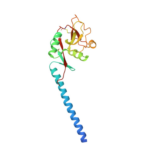
Trimeric structure of langerin.

Feinberg, H.Powlesland, A.S.Taylor, M.E.Weis, W.I.

(2010) J Biol Chem 285: 13285-13293

  • DOI: https://doi.org/10.1074/jbc.M109.086058
  • Primary Citation of Related Structures:  
    3KQG

  • PubMed Abstract: 

    Langerin, an endocytic receptor of Langerhans cells, binds pathogens such as human immunodeficiency virus by recognition of surface glycoconjugates and mediates their internalization into Birbeck granules. Langerin has an extracellular region consisting of a C-type carbohydrate-recognition domain (CRD) and a neck region that stabilizes formation of trimers. As in many other C-type lectins, oligomerization is required for high affinity binding to glycan ligands and is also likely to be important for determining specificity. To facilitate structural analysis of the human langerin trimer, a truncated form of the extracellular region, consisting of part of the neck and the CRD, has been characterized. Like the full-length protein, truncated langerin exists as a stable trimer in solution. Glycan array screening with the trimeric fragment shows that high mannose oligosaccharides are the best ligands for langerin. Structural analysis of the trimeric fragment of langerin confirms that the neck region forms a coiled-coil of alpha-helices. Multiple interactions between the neck region and the CRDs make the trimer a rigid unit with the three CRDs in fixed positions and the primary sugar-binding sites separated by a distance of 42 A. The fixed orientation of the sugar-binding sites in the trimer is likely to place constraints on the ligands that can be bound by langerin.


  • Organizational Affiliation

    Department of Structural Biology, Stanford University School of Medicine, Stanford, California 94306, USA.


Macromolecules
Find similar proteins by:  (by identity cutoff)  |  3D Structure
Entity ID: 1
MoleculeChains Sequence LengthOrganismDetailsImage
C-type lectin domain family 4 member K
A, B, C, D, E
A, B, C, D, E, F
182Homo sapiensMutation(s): 2 
Gene Names: CD207CLEC4K
UniProt & NIH Common Fund Data Resources
Find proteins for Q9UJ71 (Homo sapiens)
Explore Q9UJ71 
Go to UniProtKB:  Q9UJ71
PHAROS:  Q9UJ71
GTEx:  ENSG00000116031 
Entity Groups  
Sequence Clusters30% Identity50% Identity70% Identity90% Identity95% Identity100% Identity
UniProt GroupQ9UJ71
Sequence Annotations
Expand
  • Reference Sequence
Experimental Data & Validation

Experimental Data

  • Method: X-RAY DIFFRACTION
  • Resolution: 2.30 Å
  • R-Value Free: 0.238 
  • R-Value Work: 0.182 
  • R-Value Observed: 0.185 
  • Space Group: P 1 21 1
Unit Cell:
Length ( Å )Angle ( ˚ )
a = 80.78α = 90
b = 77.46β = 94.42
c = 85.92γ = 90
Software Package:
Software NamePurpose
Blu-Icedata collection
COMOphasing
PHENIXrefinement
MOSFLMdata reduction
SCALAdata scaling

Structure Validation

View Full Validation Report


Entry History 

Deposition Data

Revision History  (Full details and data files)

  • Version 1.0: 2010-02-23
    Type: Initial release
  • Version 1.1: 2011-07-13
    Changes: Version format compliance
  • Version 1.2: 2017-11-01
    Changes: Refinement description
  • Version 1.3: 2021-10-13
    Changes: Database references, Derived calculations
  • Version 1.4: 2024-04-03
    Changes: Data collection, Refinement description