3KP9

Structure of a bacterial homolog of vitamin K epoxide reductase


Experimental Data Snapshot

  • Method: X-RAY DIFFRACTION
  • Resolution: 3.60 Å
  • R-Value Free: 0.305 
  • R-Value Work: 0.251 
  • R-Value Observed: 0.256 

wwPDB Validation   3D Report Full Report


Ligand Structure Quality Assessment 


This is version 1.2 of the entry. See complete history


Literature

Structure of a bacterial homologue of vitamin K epoxide reductase.

Li, W.Schulman, S.Dutton, R.J.Boyd, D.Beckwith, J.Rapoport, T.A.

(2010) Nature 463: 507-512

  • DOI: https://doi.org/10.1038/nature08720
  • Primary Citation of Related Structures:  
    3KP8, 3KP9

  • PubMed Abstract: 

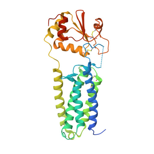
    Vitamin K epoxide reductase (VKOR) generates vitamin K hydroquinone to sustain gamma-carboxylation of many blood coagulation factors. Here, we report the 3.6 A crystal structure of a bacterial homologue of VKOR from Synechococcus sp. The structure shows VKOR in complex with its naturally fused redox partner, a thioredoxin-like domain, and corresponds to an arrested state of electron transfer. The catalytic core of VKOR is a four transmembrane helix bundle that surrounds a quinone, connected through an additional transmembrane segment with the periplasmic thioredoxin-like domain. We propose a pathway for how VKOR uses electrons from cysteines of newly synthesized proteins to reduce a quinone, a mechanism confirmed by in vitro reconstitution of vitamin K-dependent disulphide bridge formation. Our results have implications for the mechanism of the mammalian VKOR and explain how mutations can cause resistance to the VKOR inhibitor warfarin, the most commonly used oral anticoagulant.


  • Organizational Affiliation

    Howard Hughes Medical Institute and Department of Cell Biology, Harvard Medical School, 240 Longwood Avenue, Boston, Massachusetts 02115, USA. weikai@crystal.harvard.edu


Macromolecules
Find similar proteins by:  (by identity cutoff)  |  3D Structure
Entity ID: 1
MoleculeChains Sequence LengthOrganismDetailsImage
VKORC1/thioredoxin domain protein291Synechococcus sp. JA-2-3B'a(2-13)Mutation(s): 1 
Gene Names: CYB_2278
EC: 1.1.4.2
Membrane Entity: Yes 
UniProt
Find proteins for Q2JJF6 (Synechococcus sp. (strain JA-2-3B'a(2-13)))
Explore Q2JJF6 
Go to UniProtKB:  Q2JJF6
Entity Groups  
Sequence Clusters30% Identity50% Identity70% Identity90% Identity95% Identity100% Identity
UniProt GroupQ2JJF6
Sequence Annotations
Expand
  • Reference Sequence
Experimental Data & Validation

Experimental Data

  • Method: X-RAY DIFFRACTION
  • Resolution: 3.60 Å
  • R-Value Free: 0.305 
  • R-Value Work: 0.251 
  • R-Value Observed: 0.256 
  • Space Group: P 61
Unit Cell:
Length ( Å )Angle ( ˚ )
a = 136.942α = 90
b = 136.942β = 90
c = 68.48γ = 120
Software Package:
Software NamePurpose
REFMACrefinement

Structure Validation

View Full Validation Report


Ligand Structure Quality Assessment 


Entry History 

Deposition Data

Revision History  (Full details and data files)

  • Version 1.0: 2010-02-09
    Type: Initial release
  • Version 1.1: 2011-07-13
    Changes: Advisory, Version format compliance
  • Version 1.2: 2021-10-13
    Changes: Database references, Derived calculations