3KMF

Room Temperature Time-of-Flight Neutron Diffraction Study of Deoxy Human Normal Adult Hemoglobin


Experimental Data Snapshot

  • Method: NEUTRON DIFFRACTION
  • Resolution: 2.00 Å
  • R-Value Free: 0.300 
  • R-Value Work: 0.250 

wwPDB Validation   3D Report Full Report


This is version 2.1 of the entry. See complete history


Literature

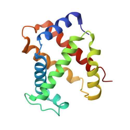
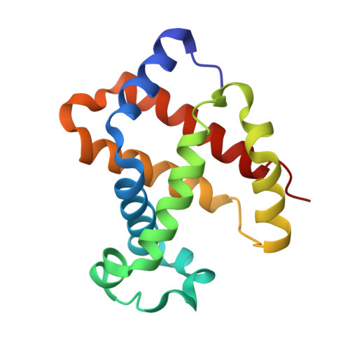
Direct Determination of Protonation States of Histidine Residues in a 2 A Neutron Structure of Deoxy-Human Normal Adult Hemoglobin and Implications for the Bohr Effect.

Kovalevsky, A.Y.Chatake, T.Shibayama, N.Park, S.Y.Ishikawa, T.Mustyakimov, M.Fisher, Z.Langan, P.Morimoto, Y.

(2010) J Mol Biol 398: 276-291

  • DOI: https://doi.org/10.1016/j.jmb.2010.03.016
  • Primary Citation of Related Structures:  
    3KMF

  • PubMed Abstract: 

    We have investigated the protonation states of histidine residues (potential Bohr groups) in the deoxy form (T state) of human hemoglobin by direct determination of hydrogen (deuterium) positions with the neutron protein crystallography technique. The reversible binding of protons is key to the allosteric regulation of human hemoglobin. The protonation states of 35 of the 38 His residues were directly determined from neutron scattering omit maps, with 3 of the remaining residues being disordered. Protonation states of 5 equivalent His residues--alpha His20, alpha His50, alpha His89, beta His143, and beta His146--differ between the symmetry-related globin subunits. The distal His residues, alpha His58 and beta His63, are protonated in the alpha 1 beta 1 heterodimer and are neutral in alpha 2 beta 2. Buried residue alpha His103 is found to be protonated in both subunits. These distal and buried residues have the potential to act as Bohr groups. The observed protonation states of His residues are compared to changes in their pK(a) values during the transition from the T to the R state and the results provide some new insights into our understanding of the molecular mechanism of the Bohr effect.


  • Organizational Affiliation

    Bioscience Division, Los Alamos National Laboratory, Los Alamos, NM 87545, USA. ayk@lanl.gov


Macromolecules
Find similar proteins by:  (by identity cutoff)  |  3D Structure
Entity ID: 1
MoleculeChains Sequence LengthOrganismDetailsImage
Hemoglobin subunit alphaA,
C [auth E]
141Homo sapiensMutation(s): 0 
UniProt & NIH Common Fund Data Resources
Find proteins for P69905 (Homo sapiens)
Explore P69905 
Go to UniProtKB:  P69905
PHAROS:  P69905
Entity Groups  
Sequence Clusters30% Identity50% Identity70% Identity90% Identity95% Identity100% Identity
UniProt GroupP69905
Sequence Annotations
Expand
  • Reference Sequence
Find similar proteins by:  (by identity cutoff)  |  3D Structure
Entity ID: 2
MoleculeChains Sequence LengthOrganismDetailsImage
Hemoglobin subunit betaB [auth C],
D [auth G]
146Homo sapiensMutation(s): 0 
UniProt & NIH Common Fund Data Resources
Find proteins for P68871 (Homo sapiens)
Explore P68871 
Go to UniProtKB:  P68871
PHAROS:  P68871
GTEx:  ENSG00000244734 
Entity Groups  
Sequence Clusters30% Identity50% Identity70% Identity90% Identity95% Identity100% Identity
UniProt GroupP68871
Sequence Annotations
Expand
  • Reference Sequence
Experimental Data & Validation

Experimental Data

  • Method: NEUTRON DIFFRACTION
  • Resolution: 2.00 Å
  • R-Value Free: 0.300 
  • R-Value Work: 0.250 
  • Space Group: P 1 21 1
Unit Cell:
Length ( Å )Angle ( ˚ )
a = 63.79α = 90
b = 84.45β = 99.33
c = 54.35γ = 90
Software Package:
Software NamePurpose
PCSdata collection
nCNSrefinement
d*TREKdata reduction
LAUENORMdata scaling
nCNSphasing

Structure Validation

View Full Validation Report


Entry History 

Deposition Data

Revision History  (Full details and data files)

  • Version 1.0: 2010-04-21
    Type: Initial release
  • Version 1.1: 2011-07-13
    Changes: Version format compliance
  • Version 2.0: 2018-06-13
    Changes: Atomic model, Data collection
  • Version 2.1: 2023-11-01
    Changes: Data collection, Database references, Derived calculations, Refinement description