3KJS

Crystal Structure of T. cruzi DHFR-TS with 3 high affinity DHFR inhibitors: DQ1 inhibitor complex


Experimental Data Snapshot

  • Method: X-RAY DIFFRACTION
  • Resolution: 2.50 Å
  • R-Value Free: 0.234 
  • R-Value Work: 0.200 
  • R-Value Observed: 0.202 

wwPDB Validation   3D Report Full Report


Ligand Structure Quality Assessment 


This is version 1.2 of the entry. See complete history


Literature

Synthesis and characterization of potent inhibitors of Trypanosoma cruzi dihydrofolate reductase.

Schormann, N.Velu, S.E.Murugesan, S.Senkovich, O.Walker, K.Chenna, B.C.Shinkre, B.Desai, A.Chattopadhyay, D.

(2010) Bioorg Med Chem 18: 4056-4066

  • DOI: https://doi.org/10.1016/j.bmc.2010.04.020
  • Primary Citation of Related Structures:  
    3KJS

  • PubMed Abstract: 

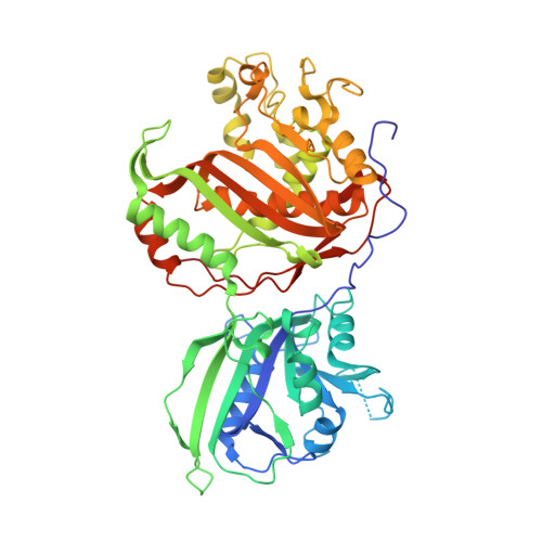
    Dihydrofolate reductase (DHFR) of the parasite Trypanosoma cruzi (T. cruzi) is a potential target for developing drugs to treat Chagas' disease. We have undertaken a detailed structure-activity study of this enzyme. We report here synthesis and characterization of six potent inhibitors of the parasitic enzyme. Inhibitory activity of each compound was determined against T. cruzi and human DHFR. One of these compounds, ethyl 4-(5-[(2,4-diamino-6-quinazolinyl)methyl]amino-2-methoxyphenoxy)butanoate (6b) was co-crystallized with the bifunctional dihydrofolate reductase-thymidylate synthase enzyme of T. cruzi and the crystal structure of the ternary enzyme:cofactor:inhibitor complex was determined. Molecular docking was used to analyze the potential interactions of all inhibitors with T. cruzi DHFR and human DHFR. Inhibitory activities of these compounds are discussed in the light of enzyme-ligand interactions. Binding affinities of each inhibitor for the respective enzymes were calculated based on the experimental or docked binding mode. An estimated 60-70% of the total binding energy is contributed by the 2,4-diaminoquinazoline scaffold.


  • Organizational Affiliation

    Department of Medicine, University of Alabama, Birmingham, Birmingham, AL 35294, USA.


Macromolecules
Find similar proteins by:  (by identity cutoff)  |  3D Structure
Entity ID: 1
MoleculeChains Sequence LengthOrganismDetailsImage
Dihydrofolate reductase-thymidylate synthase
A, B, C, D
521Trypanosoma cruziMutation(s): 0 
Gene Names: dhfrts
EC: 1.5.1.3 (PDB Primary Data), 2.1.1.45 (PDB Primary Data)
UniProt
Find proteins for Q8T5T8 (Trypanosoma cruzi)
Explore Q8T5T8 
Go to UniProtKB:  Q8T5T8
Entity Groups  
Sequence Clusters30% Identity50% Identity70% Identity90% Identity95% Identity100% Identity
UniProt GroupQ8T5T8
Sequence Annotations
Expand
  • Reference Sequence
Small Molecules
Ligands 4 Unique
IDChains Name / Formula / InChI Key2D Diagram3D Interactions
NAP
Query on NAP

Download Ideal Coordinates CCD File 
CA [auth D],
F [auth A],
N [auth B],
V [auth C]
NADP NICOTINAMIDE-ADENINE-DINUCLEOTIDE PHOSPHATE
C21 H28 N7 O17 P3
XJLXINKUBYWONI-NNYOXOHSSA-N
DQ1
Query on DQ1

Download Ideal Coordinates CCD File 
AA [auth D],
E [auth A],
M [auth B],
S [auth C]
ethyl 4-(5-{[(2,4-diaminoquinazolin-6-yl)methyl]amino}-2-methoxyphenoxy)butanoate
C22 H27 N5 O4
YUHXPHNBJFUHSN-UHFFFAOYSA-N
SO4
Query on SO4

Download Ideal Coordinates CCD File 
BA [auth D]
G [auth A]
L [auth B]
O [auth B]
T [auth C]
BA [auth D],
G [auth A],
L [auth B],
O [auth B],
T [auth C],
U [auth C],
Y [auth D],
Z [auth D]
SULFATE ION
O4 S
QAOWNCQODCNURD-UHFFFAOYSA-L
EDO
Query on EDO

Download Ideal Coordinates CCD File 
DA [auth D]
EA [auth D]
FA [auth D]
GA [auth D]
H [auth A]
DA [auth D],
EA [auth D],
FA [auth D],
GA [auth D],
H [auth A],
I [auth A],
J [auth A],
K [auth A],
P [auth B],
Q [auth B],
R [auth B],
W [auth C],
X [auth C]
1,2-ETHANEDIOL
C2 H6 O2
LYCAIKOWRPUZTN-UHFFFAOYSA-N
Binding Affinity Annotations 
IDSourceBinding Affinity
DQ1 BindingDB:  3KJS Ki: min: 1.3, max: 5.68 (nM) from 2 assay(s)
IC50: 23.8 (nM) from 1 assay(s)
Binding MOAD:  3KJS Ki: 1.3 (nM) from 1 assay(s)
Experimental Data & Validation

Experimental Data

  • Method: X-RAY DIFFRACTION
  • Resolution: 2.50 Å
  • R-Value Free: 0.234 
  • R-Value Work: 0.200 
  • R-Value Observed: 0.202 
  • Space Group: P 43 21 2
Unit Cell:
Length ( Å )Angle ( ˚ )
a = 175.62α = 90
b = 175.62β = 90
c = 249.905γ = 90
Software Package:
Software NamePurpose
HKL-2000data collection
PHASERphasing
REFMACrefinement
d*TREKdata reduction
d*TREKdata scaling

Structure Validation

View Full Validation Report


Ligand Structure Quality Assessment 


Entry History 

Deposition Data

Revision History  (Full details and data files)

  • Version 1.0: 2010-06-09
    Type: Initial release
  • Version 1.1: 2011-07-13
    Changes: Version format compliance
  • Version 1.2: 2023-09-06
    Changes: Data collection, Database references, Derived calculations, Refinement description