3KIJ

Crystal structure of the human PDI-peroxidase


Experimental Data Snapshot

  • Method: X-RAY DIFFRACTION
  • Resolution: 1.80 Å
  • R-Value Free: 0.248 
  • R-Value Work: 0.209 
  • R-Value Observed: 0.211 

wwPDB Validation   3D Report Full Report


This is version 1.3 of the entry. See complete history


Literature

Two Endoplasmic Reticulum PDI Peroxidases Increase the Efficiency of the Use of Peroxide during Disulfide Bond Formation.

Nguyen, V.D.Saaranen, M.J.Karala, A.R.Lappi, A.K.Wang, L.Raykhel, I.B.Alanen, H.I.Salo, K.E.Wang, C.C.Ruddock, L.W.

(2011) J Mol Biol 406: 503-515

  • DOI: https://doi.org/10.1016/j.jmb.2010.12.039
  • Primary Citation of Related Structures:  
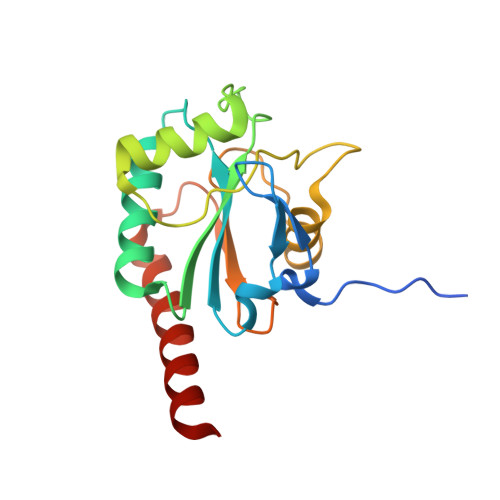
    3KIJ

  • PubMed Abstract: 

    Disulfide bond formation in the endoplasmic reticulum by the sulfhydryl oxidase Ero1 family is thought to be accompanied by the concomitant formation of hydrogen peroxide. Since secretory cells can make substantial amounts of proteins that contain disulfide bonds, the production of this reactive oxygen species could have potentially lethal consequences. Here, we show that two human proteins, GPx7 and GPx8, labeled as secreted glutathione peroxidases, are actually endoplasmic reticulum-resident protein disulfide isomerase peroxidases. In vitro, the addition of GPx7 or GPx8 to a folding protein along with protein disulfide isomerase and peroxide enables the efficient oxidative refolding of a reduced denatured protein. Furthermore, both GPx7 and GPx8 interact with Ero1α in vivo, and GPx7 significantly increases oxygen consumption by Ero1α in vitro. Hence, GPx7 and GPx8 may represent a novel route for the productive use of peroxide produced by Ero1α during disulfide bond formation.


  • Organizational Affiliation

    Department of Biochemistry, University of Oulu, Linnanmaa Campus, 90570 Oulu, Finland.


Macromolecules
Find similar proteins by:  (by identity cutoff)  |  3D Structure
Entity ID: 1
MoleculeChains Sequence LengthOrganismDetailsImage
Probable glutathione peroxidase 8
A, B, C
180Homo sapiensMutation(s): 0 
Gene Names: GPX8GPX8_humanUNQ847/PRO1785
EC: 1.11.1.9
Membrane Entity: Yes 
UniProt & NIH Common Fund Data Resources
Find proteins for Q8TED1 (Homo sapiens)
Explore Q8TED1 
Go to UniProtKB:  Q8TED1
PHAROS:  Q8TED1
GTEx:  ENSG00000164294 
Entity Groups  
Sequence Clusters30% Identity50% Identity70% Identity90% Identity95% Identity100% Identity
UniProt GroupQ8TED1
Sequence Annotations
Expand
  • Reference Sequence
Small Molecules
Ligands 1 Unique
IDChains Name / Formula / InChI Key2D Diagram3D Interactions
SO4
Query on SO4

Download Ideal Coordinates CCD File 
D [auth B]SULFATE ION
O4 S
QAOWNCQODCNURD-UHFFFAOYSA-L
Experimental Data & Validation

Experimental Data

  • Method: X-RAY DIFFRACTION
  • Resolution: 1.80 Å
  • R-Value Free: 0.248 
  • R-Value Work: 0.209 
  • R-Value Observed: 0.211 
  • Space Group: C 1 2 1
Unit Cell:
Length ( Å )Angle ( ˚ )
a = 90.43α = 90
b = 122.3β = 117.9
c = 70.18γ = 90
Software Package:
Software NamePurpose
MAR345data collection
PHASERphasing
REFMACrefinement
XDSdata reduction
XSCALEdata scaling

Structure Validation

View Full Validation Report


Entry History 

Deposition Data

Revision History  (Full details and data files)

  • Version 1.0: 2011-01-12
    Type: Initial release
  • Version 1.1: 2011-07-13
    Changes: Version format compliance
  • Version 1.2: 2017-11-01
    Changes: Refinement description
  • Version 1.3: 2023-09-06
    Changes: Data collection, Database references, Derived calculations, Refinement description