3KHC

Crystal Structure of Escherichia coli AlkB in complex with ssDNA containing a 1-methylguanine lesion


Experimental Data Snapshot

  • Method: X-RAY DIFFRACTION
  • Resolution: 2.20 Å
  • R-Value Free: 0.273 
  • R-Value Work: 0.211 
  • R-Value Observed: 0.214 

wwPDB Validation   3D Report Full Report


This is version 1.3 of the entry. See complete history


Literature

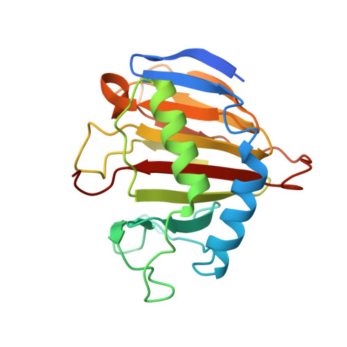
Structural and mutational analysis of Escherichia coli AlkB provides insight into substrate specificity and DNA damage searching.

Holland, P.J.Hollis, T.

(2010) PLoS One 5: e8680-e8680

  • DOI: https://doi.org/10.1371/journal.pone.0008680
  • Primary Citation of Related Structures:  
    3KHB, 3KHC

  • PubMed Abstract: 

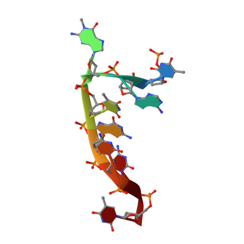
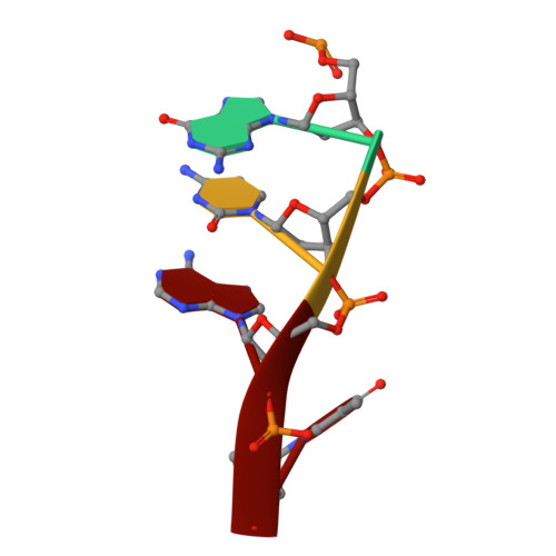
    In Escherichia coli, cytotoxic DNA methyl lesions on the N1 position of purines and N3 position of pyrimidines are primarily repaired by the 2-oxoglutarate (2-OG) iron(II) dependent dioxygenase, AlkB. AlkB repairs 1-methyladenine (1-meA) and 3-methylcytosine (3-meC) lesions, but it also repairs 1-methylguanine (1-meG) and 3-methylthymine (3-meT) at a much less efficient rate. How the AlkB enzyme is able to locate and identify methylated bases in ssDNA has remained an open question.


  • Organizational Affiliation

    Department of Biochemistry, Wake Forest University School of Medicine, Winston-Salem, North Carolina, USA.


Macromolecules

Find similar proteins by:  (by identity cutoff)  |  3D Structure
Entity ID: 1
MoleculeChains Sequence LengthOrganismDetailsImage
Alpha-ketoglutarate-dependent dioxygenase alkB
A, B
219Escherichia coli K-12Mutation(s): 1 
Gene Names: aidDalkBb2212JW2200
EC: 1.14.11
UniProt
Find proteins for P05050 (Escherichia coli (strain K12))
Explore P05050 
Go to UniProtKB:  P05050
Entity Groups  
Sequence Clusters30% Identity50% Identity70% Identity90% Identity95% Identity100% Identity
UniProt GroupP05050
Sequence Annotations
Expand
  • Reference Sequence

Find similar nucleic acids by:  Sequence   |   3D Structure  

Entity ID: 2
MoleculeChains LengthOrganismImage
DNA (5'-D(P*TP*AP*(MG1)P*TP*GP*CP*CP*T)-3')8N/A
Sequence Annotations
Expand
  • Reference Sequence

Find similar nucleic acids by:  Sequence   |   3D Structure  

Entity ID: 3
MoleculeChains LengthOrganismImage
DNA (5'-D(P*GP*CP*AP*T)-3')4N/A
Sequence Annotations
Expand
  • Reference Sequence
Experimental Data & Validation

Experimental Data

  • Method: X-RAY DIFFRACTION
  • Resolution: 2.20 Å
  • R-Value Free: 0.273 
  • R-Value Work: 0.211 
  • R-Value Observed: 0.214 
  • Space Group: C 1 2 1
Unit Cell:
Length ( Å )Angle ( ˚ )
a = 147.809α = 90
b = 41.441β = 120.98
c = 85.622γ = 90
Software Package:
Software NamePurpose
CrystalCleardata collection
PHASERphasing
REFMACrefinement
d*TREKdata reduction
d*TREKdata scaling

Structure Validation

View Full Validation Report


Entry History 

Deposition Data

Revision History  (Full details and data files)

  • Version 1.0: 2010-01-12
    Type: Initial release
  • Version 1.1: 2011-07-13
    Changes: Advisory, Version format compliance
  • Version 1.2: 2021-10-13
    Changes: Database references, Derived calculations
  • Version 1.3: 2023-09-06
    Changes: Data collection, Refinement description