3KG6

Dehydratase domain from CurF module of Curacin polyketide synthase


Experimental Data Snapshot

  • Method: X-RAY DIFFRACTION
  • Resolution: 2.70 Å
  • R-Value Free: 0.265 
  • R-Value Work: 0.213 
  • R-Value Observed: 0.215 

wwPDB Validation   3D Report Full Report


This is version 1.4 of the entry. See complete history


Literature

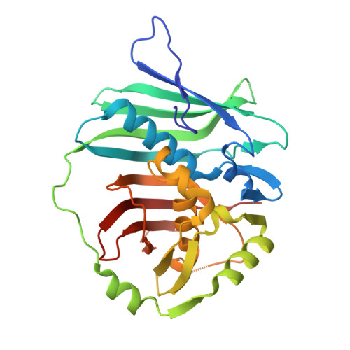
Crystal Structures of Dehydratase Domains from the Curacin Polyketide Biosynthetic Pathway.

Akey, D.L.Razelun, J.R.Tehranisa, J.Sherman, D.H.Gerwick, W.H.Smith, J.L.

(2010) Structure 18: 94-105

  • DOI: https://doi.org/10.1016/j.str.2009.10.018
  • Primary Citation of Related Structures:  
    3KG6, 3KG7, 3KG8, 3KG9

  • PubMed Abstract: 

    Modular polyketide synthases (PKS) make novel natural products through a series of preprogrammed chemical steps catalyzed by an assembly line of multidomain modules. Each assembly-line step involves unique extension and modification reactions, resulting in tremendous diversity of polyketide products. Dehydratase domains catalyze formation of an alpha,beta-double bond in the nascent polyketide intermediate. We present crystal structures of the four dehydratase domains from the curacin A PKS. The catalytic residues and substrate binding site reside in a tunnel within a single monomer. The positions of the catalytic residues and shape of the substrate tunnel explain how chirality of the substrate hydroxyl group may determine the configuration of the product double bond. Access to the active site may require opening the substrate tunnel, forming an open trench. The arrangement of monomers within the dimer is consistent among PKS dehydratases and differs from that seen in the related mammalian fatty acid synthases.


  • Organizational Affiliation

    Life Sciences Institute and Department of Biological Chemistry, University of Michigan, Ann Arbor, MI 48109, USA.


Macromolecules
Find similar proteins by:  (by identity cutoff)  |  3D Structure
Entity ID: 1
MoleculeChains Sequence LengthOrganismDetailsImage
CurF
A, B, C, D
285Lyngbya majusculaMutation(s): 0 
Gene Names: curF
EC: 4.2.1.61
UniProt
Find proteins for Q6DNE7 (Lyngbya majuscula)
Explore Q6DNE7 
Go to UniProtKB:  Q6DNE7
Entity Groups  
Sequence Clusters30% Identity50% Identity70% Identity90% Identity95% Identity100% Identity
UniProt GroupQ6DNE7
Sequence Annotations
Expand
  • Reference Sequence
Experimental Data & Validation

Experimental Data

  • Method: X-RAY DIFFRACTION
  • Resolution: 2.70 Å
  • R-Value Free: 0.265 
  • R-Value Work: 0.213 
  • R-Value Observed: 0.215 
  • Space Group: C 2 2 21
Unit Cell:
Length ( Å )Angle ( ˚ )
a = 87.379α = 90
b = 198.385β = 90
c = 146.764γ = 90
Software Package:
Software NamePurpose
DENZOdata reduction
SCALEPACKdata scaling
PHASERphasing
RESOLVEphasing
REFMACrefinement
PDB_EXTRACTdata extraction
HKL-2000data reduction
HKL-2000data scaling

Structure Validation

View Full Validation Report


Entry History 

Deposition Data

Revision History  (Full details and data files)

  • Version 1.0: 2010-01-19
    Type: Initial release
  • Version 1.1: 2011-07-13
    Changes: Advisory, Refinement description, Version format compliance
  • Version 1.2: 2017-11-01
    Changes: Refinement description
  • Version 1.3: 2023-09-06
    Changes: Data collection, Database references, Derived calculations, Refinement description
  • Version 1.4: 2024-01-31
    Changes: Refinement description