3KEG

X-ray Crystallographic Structure of a Y131F mutant of Pseudomonas Aeruginosa Azoreductase in complex with Methyl RED


Experimental Data Snapshot

  • Method: X-RAY DIFFRACTION
  • Resolution: 2.10 Å
  • R-Value Free: 0.210 
  • R-Value Work: 0.182 
  • R-Value Observed: 0.184 

wwPDB Validation   3D Report Full Report


Ligand Structure Quality Assessment 


This is version 1.3 of the entry. See complete history


Literature

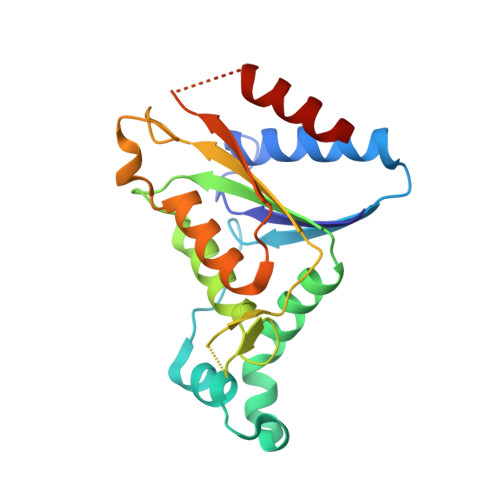
Role of tyrosine 131 in the active site of paAzoR1, an azoreductase with specificity for the inflammatory bowel disease prodrug balsalazide

Wang, C.-J.Laurieri, N.Abuhammad, A.Lowe, E.Westwood, I.Ryan, A.Sim, E.

(2010) Acta Crystallogr Sect F Struct Biol Cryst Commun 66: 2-7

  • DOI: https://doi.org/10.1107/S1744309109044741
  • Primary Citation of Related Structures:  
    3KEG

  • PubMed Abstract: 

    Azoreductase 1 from Pseudomonas aeruginosa strain PAO1 (paAzoR1) catalyses the activation of the prodrug balsalazide and reduces the azo dye methyl red using reduced nicotinamide adenine dinucleotide cofactor as an electron donor. To investigate the mechanism of the enzyme, a Y131F mutation was introduced and the enzymic properties of the mutant were compared with those of the wild-type enzyme. The crystallographic structure of the mutant with methyl red bound was solved at 2.1 A resolution and compared with the wild-type structure. Tyr131 is important in the architecture of the active site but is not essential for enzymic activity.


  • Organizational Affiliation

    Department of Pharmacology, University of Oxford, Mansfield Road, Oxford OX1 3QT, England.


Macromolecules
Find similar proteins by:  (by identity cutoff)  |  3D Structure
Entity ID: 1
MoleculeChains Sequence LengthOrganismDetailsImage
FMN-dependent NADH-azoreductase 1
A, B
212Pseudomonas aeruginosa PAO1Mutation(s): 1 
Gene Names: PA0785
EC: 1.7
UniProt
Find proteins for Q9I5F3 (Pseudomonas aeruginosa (strain ATCC 15692 / DSM 22644 / CIP 104116 / JCM 14847 / LMG 12228 / 1C / PRS 101 / PAO1))
Explore Q9I5F3 
Go to UniProtKB:  Q9I5F3
Entity Groups  
Sequence Clusters30% Identity50% Identity70% Identity90% Identity95% Identity100% Identity
UniProt GroupQ9I5F3
Sequence Annotations
Expand
  • Reference Sequence
Experimental Data & Validation

Experimental Data

  • Method: X-RAY DIFFRACTION
  • Resolution: 2.10 Å
  • R-Value Free: 0.210 
  • R-Value Work: 0.182 
  • R-Value Observed: 0.184 
  • Space Group: P 31 2 1
Unit Cell:
Length ( Å )Angle ( ˚ )
a = 82.81α = 90
b = 82.81β = 90
c = 108.91γ = 120
Software Package:
Software NamePurpose
MOSFLMdata reduction
SCALAdata scaling
PHASERphasing
PHENIXrefinement
PDB_EXTRACTdata extraction

Structure Validation

View Full Validation Report


Ligand Structure Quality Assessment 


Entry History 

Deposition Data

Revision History  (Full details and data files)

  • Version 1.0: 2010-01-12
    Type: Initial release
  • Version 1.1: 2011-07-13
    Changes: Version format compliance
  • Version 1.2: 2021-11-10
    Changes: Database references, Derived calculations
  • Version 1.3: 2023-11-01
    Changes: Data collection, Refinement description