


The crystal structure of the RsbU and RsbW domains of Rv1364c from Mycobacterium tuberculosis
King-Scott, J., Panjikar, S., Tucker, P.A.To be published.
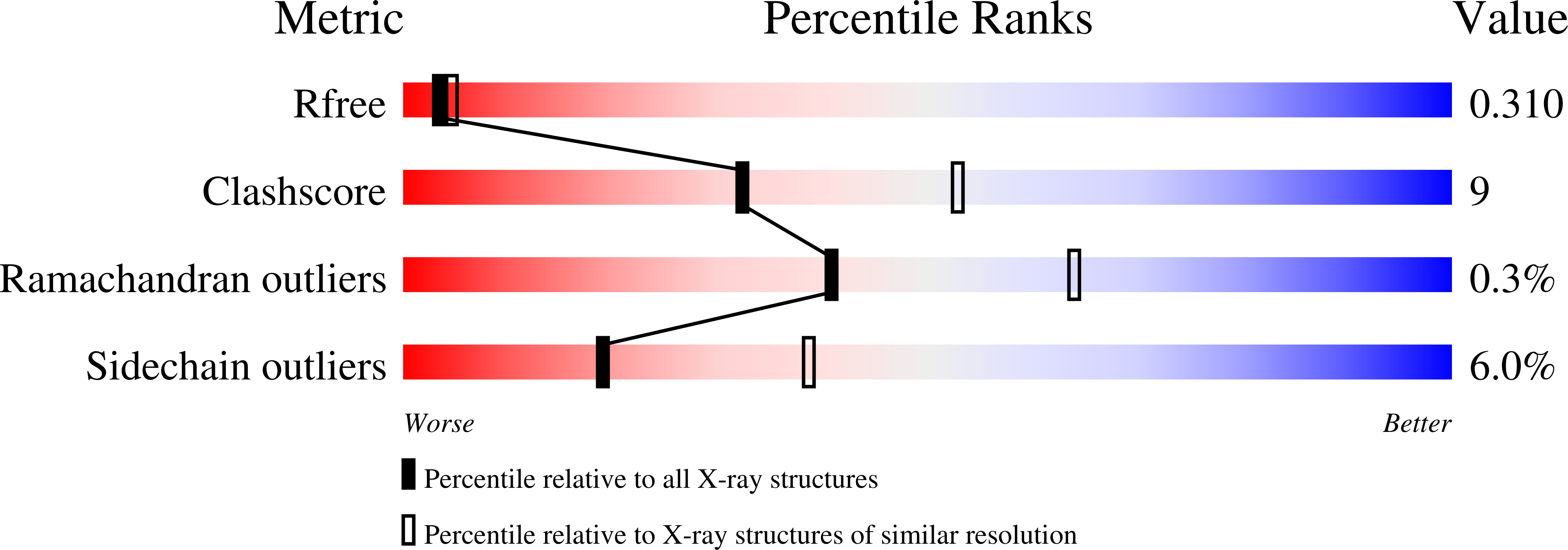
Experimental Data Snapshot
wwPDB Validation 3D Report Full Report
Entity ID: 1 | |||||
|---|---|---|---|---|---|
| Molecule | Chains | Sequence Length | Organism | Details | Image |
| Protein Rv1364c/MT1410 | 399 | Mycobacterium tuberculosis | Mutation(s): 0 Gene Names: MT1410, MTCY02B10.28c, Rv1364c | 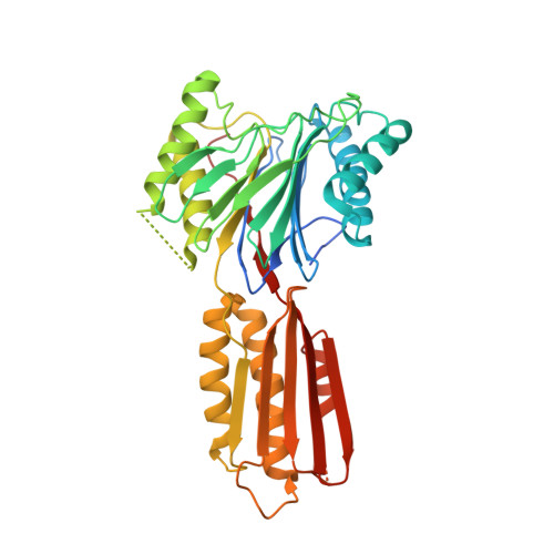 | |
UniProt | |||||
Find proteins for P9WLZ7 (Mycobacterium tuberculosis (strain ATCC 25618 / H37Rv)) Explore P9WLZ7 Go to UniProtKB: P9WLZ7 | |||||
Entity Groups | |||||
| Sequence Clusters | 30% Identity50% Identity70% Identity90% Identity95% Identity100% Identity | ||||
| UniProt Group | P9WLZ7 | ||||
Sequence AnnotationsExpand | |||||
| |||||
| Ligands 3 Unique | |||||
|---|---|---|---|---|---|
| ID | Chains | Name / Formula / InChI Key | 2D Diagram | 3D Interactions | |
| SO4 Query on SO4 | C [auth A] | SULFATE ION O4 S QAOWNCQODCNURD-UHFFFAOYSA-L |  | ||
| GOL Query on GOL | D [auth A] | GLYCEROL C3 H8 O3 PEDCQBHIVMGVHV-UHFFFAOYSA-N |  | ||
| MN Query on MN | E [auth A], F [auth A], G [auth B], H [auth B] | MANGANESE (II) ION Mn WAEMQWOKJMHJLA-UHFFFAOYSA-N |  | ||
| Length ( Å ) | Angle ( ˚ ) |
|---|---|
| a = 100.118 | α = 90 |
| b = 100.118 | β = 90 |
| c = 169.653 | γ = 90 |
| Software Name | Purpose |
|---|---|
| MAR345dtb | data collection |
| MOLREP | phasing |
| REFMAC | refinement |
| DENZO | data reduction |
| SCALEPACK | data scaling |
RCSB PDB (citation) is hosted by
RCSB PDB is a member of the

