3KD7

Designed TPR module (CTPR390) in complex with its peptide-ligand (Hsp90 peptide)


Experimental Data Snapshot

  • Method: X-RAY DIFFRACTION
  • Resolution: 2.85 Å
  • R-Value Free: 0.283 
  • R-Value Work: 0.271 
  • R-Value Observed: 0.271 

wwPDB Validation   3D Report Full Report


This is version 1.2 of the entry. See complete history


Literature

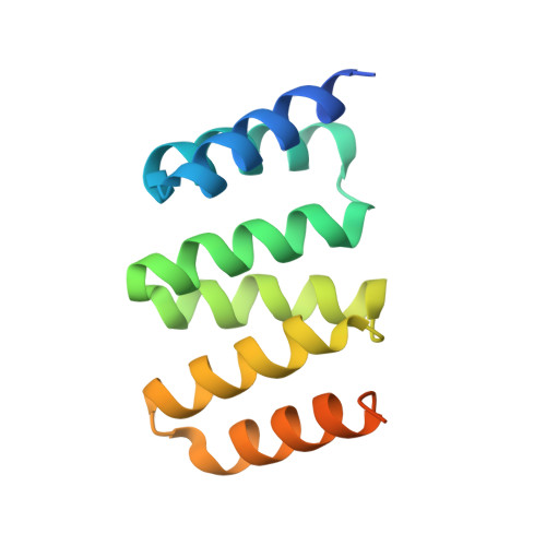
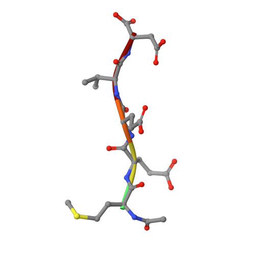
Crystal structure of a designed tetratricopeptide repeat module in complex with its peptide ligand.

Cortajarena, A.L.Wang, J.Regan, L.

(2010) FEBS J 277: 1058-1066

  • DOI: https://doi.org/10.1111/j.1742-4658.2009.07549.x
  • Primary Citation of Related Structures:  
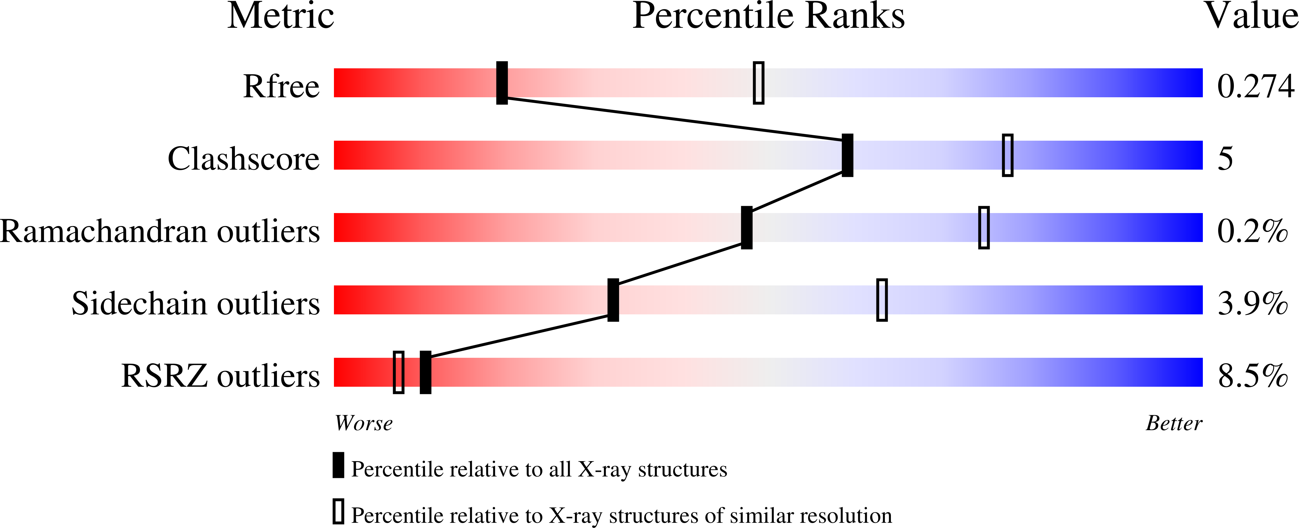
    3KD7

  • PubMed Abstract: 

    Tetratricopeptide repeats (TPRs) are protein domains that mediate key protein-protein interactions in cells. Several TPR domains bind the C-termini of the chaperones heat shock protein (Hsp)90 and/or Hsp70, and exchange of such binding partners is key for the heat shock response. We have previously described the design of a TPR protein that binds tightly and specifically to the C-terminus of Hsp90, and in doing so, is able to inhibit chaperone function in vivo. Here we present the X-ray crystal structure of the designed TPR domain (CTPR390) in complex with its peptide ligand--the C-terminal residues of Hsp90 (peptide MEEVD). This structure reveals two interesting aspects of the TPR modules. First, a new packing arrangement of 3-TPR modules is observed. The TPR units stack against each other in an unusual fashion to form infinite superhelices in the crystal. Second, the structure provides insights into the molecular basis of TPR-ligand recognition.


  • Organizational Affiliation

    Department of Molecular Biophysics & Biochemistry, Yale University, New Haven, CT, USA.


Macromolecules
Find similar proteins by:  (by identity cutoff)  |  3D Structure
Entity ID: 1
MoleculeChains Sequence LengthOrganismDetailsImage
CTPR390
A, B, C, D, E
125unidentifiedMutation(s): 0 
Entity Groups  
Sequence Clusters30% Identity50% Identity70% Identity90% Identity95% Identity100% Identity
Sequence Annotations
Expand
  • Reference Sequence

Find similar proteins by:  Sequence   |   3D Structure  

Entity ID: 2
MoleculeChains Sequence LengthOrganismDetailsImage
Hsp90 MEEVD peptideF [auth G],
G [auth H],
H [auth I],
I [auth J],
J [auth K]
6N/AMutation(s): 0 
UniProt
Find proteins for Q9H2A1 (Homo sapiens)
Explore Q9H2A1 
Go to UniProtKB:  Q9H2A1
Entity Groups  
UniProt GroupQ9H2A1
Sequence Annotations
Expand
  • Reference Sequence
Experimental Data & Validation

Experimental Data

  • Method: X-RAY DIFFRACTION
  • Resolution: 2.85 Å
  • R-Value Free: 0.283 
  • R-Value Work: 0.271 
  • R-Value Observed: 0.271 
  • Space Group: H 3
Unit Cell:
Length ( Å )Angle ( ˚ )
a = 100.674α = 90
b = 100.674β = 90
c = 161.574γ = 120
Software Package:
Software NamePurpose
HKL-2000data collection
MOLREPphasing
REFMACrefinement
HKL-2000data reduction
HKL-2000data scaling

Structure Validation

View Full Validation Report


Entry History 

Deposition Data

Revision History  (Full details and data files)

  • Version 1.0: 2010-02-02
    Type: Initial release
  • Version 1.1: 2011-07-13
    Changes: Advisory, Refinement description, Version format compliance
  • Version 1.2: 2023-09-06
    Changes: Data collection, Database references, Derived calculations, Refinement description