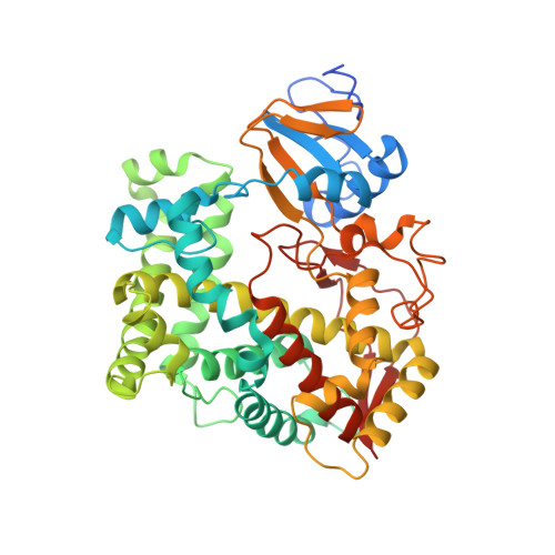
Crystal structure of CYP24A1, a mitochondrial cytochrome P450 involved in vitamin D metabolism.
Annalora, A.J., Goodin, D.B., Hong, W.X., Zhang, Q., Johnson, E.F., Stout, C.D.(2010) J Mol Biol 396: 441-451
- PubMed: 19961857
- DOI: https://doi.org/10.1016/j.jmb.2009.11.057
- Primary Citation of Related Structures:
3K9V, 3K9Y - PubMed Abstract:
Cytochrome P450 (CYP) 24A1 catalyzes the side-chain oxidation of the hormonal form of vitamin D. Expression of CYP24A1 is up-regulated to attenuate vitamin D signaling associated with calcium homeostasis and cellular growth processes. The development of therapeutics for disorders linked to vitamin D insufficiency would be greatly facilitated by structural knowledge of CYP24A1. Here, we report the crystal structure of rat CYP24A1 at 2.5 A resolution. The structure exhibits an open cleft leading to the active-site heme prosthetic group on the distal surface that is likely to define the path of substrate access into the active site. The entrance to the cleft is flanked by conserved hydrophobic residues on helices A' and G', suggesting a mode of insertion into the inner mitochondrial membrane. A docking model for 1alpha,25-dihydroxyvitamin D(3) binding in the open form of CYP24A1 that clarifies the structural determinants of secosteroid recognition and validates the predictive power of existing homology models of CYP24A1 is proposed. Analysis of CYP24A1's proximal surface identifies the determinants of adrenodoxin recognition as a constellation of conserved residues from helices K, K'', and L that converge with an adjacent lysine-rich loop for binding the redox protein. Overall, the CYP24A1 structure provides the first template for understanding membrane insertion, substrate binding, and redox partner interaction in mitochondrial P450s.
Organizational Affiliation:
Department of Molecular Biology, The Scripps Research Institute, La Jolla, CA 92037, USA. annalora@scripps.edu