3K8U

Crystal Structure of the Peptidase Domain of Streptococcus ComA, a Bi-functional ABC Transporter Involved in Quorum Sensing Pathway


Experimental Data Snapshot

  • Method: X-RAY DIFFRACTION
  • Resolution: 1.90 Å
  • R-Value Free: 0.234 
  • R-Value Work: 0.212 
  • R-Value Observed: 0.212 

wwPDB Validation   3D Report Full Report


This is version 1.3 of the entry. See complete history


Literature

Crystal structure of the peptidase domain of Streptococcus ComA, a bifunctional ATP-binding cassette transporter involved in the quorum-sensing pathway

Ishii, S.Yano, T.Ebihara, A.Okamoto, A.Manzoku, M.Hayashi, H.

(2010) J Biol Chem 285: 10777-10785

  • DOI: https://doi.org/10.1074/jbc.M109.093781
  • Primary Citation of Related Structures:  
    3K8U

  • PubMed Abstract: 

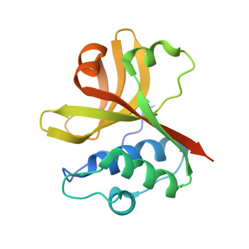
    ComA of Streptococcus is a member of the bacteriocin-associated ATP-binding cassette transporter family and is postulated to be responsible for both the processing of the propeptide ComC and secretion of the mature quorum-sensing signal. The 150-amino acid peptidase domain (PEP) of ComA specifically recognizes an extended region of ComC that is 15 amino acids in length. It has been proposed that an amphipathic alpha-helix formed by the N-terminal leader region of ComC, as well as the Gly-Gly motif at the cleavage site, is critical for the PEP-ComC interaction. To elucidate the substrate recognition mechanism, we determined the three-dimensional crystal structure of Streptococcus mutans PEP and then constructed models for the PEP.ComC complexes. PEP had an overall structure similar to the papain-like cysteine proteases as has long been predicted. The active site was located at the bottom of a narrow cleft, which is suitable for binding the Gly-Gly motif. Together with the results from mutational experiments, a shallow hydrophobic concave surface of PEP was proposed as a site that accommodates the N-terminal helix of ComC. This dual mode of substrate recognition would provide the small PEP domain with an extremely high substrate specificity.


  • Organizational Affiliation

    Department of Biochemistry, Osaka Medical College, Takatsuki, Osaka 569-8686, Japan.


Macromolecules
Find similar proteins by:  (by identity cutoff)  |  3D Structure
Entity ID: 1
MoleculeChains Sequence LengthOrganismDetailsImage
Putative ABC transporter, ATP-binding protein ComA156Streptococcus mutansMutation(s): 0 
UniProt
Find proteins for Q8DW05 (Streptococcus mutans serotype c (strain ATCC 700610 / UA159))
Explore Q8DW05 
Go to UniProtKB:  Q8DW05
Entity Groups  
Sequence Clusters30% Identity50% Identity70% Identity90% Identity95% Identity100% Identity
UniProt GroupQ8DW05
Sequence Annotations
Expand
  • Reference Sequence
Experimental Data & Validation

Experimental Data

  • Method: X-RAY DIFFRACTION
  • Resolution: 1.90 Å
  • R-Value Free: 0.234 
  • R-Value Work: 0.212 
  • R-Value Observed: 0.212 
  • Space Group: P 43 21 2
Unit Cell:
Length ( Å )Angle ( ˚ )
a = 58.61α = 90
b = 58.61β = 90
c = 112.05γ = 90
Software Package:
Software NamePurpose
HKL-2000data collection
SHELXSphasing
CNSrefinement
HKL-2000data reduction
HKL-2000data scaling

Structure Validation

View Full Validation Report


Entry History 

Deposition Data

Revision History  (Full details and data files)

  • Version 1.0: 2010-02-23
    Type: Initial release
  • Version 1.1: 2011-07-13
    Changes: Version format compliance
  • Version 1.2: 2014-02-26
    Changes: Database references
  • Version 1.3: 2024-03-20
    Changes: Data collection, Database references