3K7V

Protein phosphatase 2A core complex bound to dinophysistoxin-1


Experimental Data Snapshot

  • Method: X-RAY DIFFRACTION
  • Resolution: 2.85 Å
  • R-Value Free: 0.262 
  • R-Value Work: 0.217 
  • R-Value Observed: 0.217 

wwPDB Validation   3D Report Full Report


Ligand Structure Quality Assessment 


This is version 1.2 of the entry. See complete history


Literature

A structural basis for the reduced toxicity of dinophysistoxin-2.

Huhn, J.Jeffrey, P.D.Larsen, K.Rundberget, T.Rise, F.Cox, N.R.Arcus, V.Shi, Y.Miles, C.O.

(2009) Chem Res Toxicol 22: 1782-1786

  • DOI: https://doi.org/10.1021/tx9001622
  • Primary Citation of Related Structures:  
    3K7V, 3K7W

  • PubMed Abstract: 

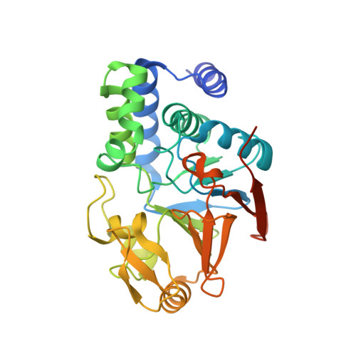
    Okadaic acid (OA), dinophysistoxin-1 (DTX-1), and dinophysistoxin-2 (DTX-2) are algal toxins that can accumulate in shellfish and cause diarrhetic shellfish poisoning. Recent studies indicate that DTX-2 is about half as toxic and has about half the affinity for protein phosphatase 2A (PP2A) as OA. NMR structural studies showed that DTX-1 possessed an equatorial 35-methyl group but that DTX-2 had an axial 35-methyl group. Molecular modeling studies indicated that an axial 35-methyl could exhibit unfavorable interactions in the PP2A binding site, and this has been proposed as the reason for the reduced toxicity of DTX-2. Statistical analyses of published data indicate that the affinity of PP2A for DTX-1 is 1.6-fold higher, and for DTX-2 is 2-fold lower, than for OA. We obtained X-ray crystal structures of DTX-1 and DTX-2 bound to PP2A. The crystal structures independently confirm the C-35 stereochemistries determined in the earlier NMR study. The structure for the DTX-1 complex was virtually identical to that of the OA-PP2A complex, except for the presence of the equatorial 35-methyl on the ligand. The favorable placement of the equatorial 35-methyl group of DTX-1 against the aromatic pi-bonds of His191 may account for the increased affinity of PP2A toward DTX-1. In contrast, the axial 35-methyl of DTX-2 caused the side chain of His191 to rotate 140 degrees so that it pointed toward the solvent, thereby opening one end of the hydrophobic binding cage. This rearrangement to accommodate the unfavorable interaction from the axial 35-methyl of DTX-2 reduces the binding energy and appears to be responsible for the reduced affinity of PP2A for DTX-2. These results highlight the potential of molecular modeling studies for understanding the relative toxicity of analogues once the binding site at the molecular target has been properly characterized.


  • Organizational Affiliation

    Department of Molecular Biology, Lewis Thomas Laboratory, Princeton University, Washington Road, Princeton, New Jersey 08544, USA.


Macromolecules
Find similar proteins by:  (by identity cutoff)  |  3D Structure
Entity ID: 1
MoleculeChains Sequence LengthOrganismDetailsImage
Serine/threonine-protein phosphatase 2A 65 kDa regulatory subunit A alpha isoform589Homo sapiensMutation(s): 0 
Gene Names: PPP2R1A
UniProt & NIH Common Fund Data Resources
Find proteins for P30153 (Homo sapiens)
Explore P30153 
Go to UniProtKB:  P30153
PHAROS:  P30153
GTEx:  ENSG00000105568 
Entity Groups  
Sequence Clusters30% Identity50% Identity70% Identity90% Identity95% Identity100% Identity
UniProt GroupP30153
Sequence Annotations
Expand
  • Reference Sequence
Find similar proteins by:  (by identity cutoff)  |  3D Structure
Entity ID: 2
MoleculeChains Sequence LengthOrganismDetailsImage
Serine/threonine-protein phosphatase 2A catalytic subunit alpha isoformB [auth C]309Homo sapiensMutation(s): 0 
Gene Names: PPP2CA
EC: 3.1.3.16
UniProt & NIH Common Fund Data Resources
Find proteins for P67775 (Homo sapiens)
Explore P67775 
Go to UniProtKB:  P67775
PHAROS:  P67775
GTEx:  ENSG00000113575 
Entity Groups  
Sequence Clusters30% Identity50% Identity70% Identity90% Identity95% Identity100% Identity
UniProt GroupP67775
Sequence Annotations
Expand
  • Reference Sequence
Small Molecules
Ligands 3 Unique
IDChains Name / Formula / InChI Key2D Diagram3D Interactions
XT1
Query on XT1

Download Ideal Coordinates CCD File 
D [auth C](2R)-3-[(2S,5R,6R,8S)-8-{(1R,2E)-3-[(2R,4a'R,5R,6'S,8'R,8a'S)-6'-{(1S,3S)-3-[(2S,3R,6R,11R)-3,11-dimethyl-1,7-dioxaspiro[5.5]undec-2-yl]-1-hydroxybutyl}-8'-hydroxy-7'-methylideneoctahydro-3H,3'H-spiro[furan-2,2'-pyrano[3,2-b]pyran]-5-yl]-1-methylprop-2-en-1-yl}-5-hydroxy-10-methyl-1,7-dioxaspiro[5.5]undec-10-en-2-yl]-2-hydroxy-2-methylpropanoic acid
C45 H70 O13
CLBIEZBAENPDFY-HNXGFDTJSA-N
SO4
Query on SO4

Download Ideal Coordinates CCD File 
C [auth A],
G [auth C],
H [auth C]
SULFATE ION
O4 S
QAOWNCQODCNURD-UHFFFAOYSA-L
MN
Query on MN

Download Ideal Coordinates CCD File 
E [auth C],
F [auth C]
MANGANESE (II) ION
Mn
WAEMQWOKJMHJLA-UHFFFAOYSA-N
Experimental Data & Validation

Experimental Data

  • Method: X-RAY DIFFRACTION
  • Resolution: 2.85 Å
  • R-Value Free: 0.262 
  • R-Value Work: 0.217 
  • R-Value Observed: 0.217 
  • Space Group: I 2 2 2
Unit Cell:
Length ( Å )Angle ( ˚ )
a = 92.83α = 90
b = 194.78β = 90
c = 202.1γ = 90
Software Package:
Software NamePurpose
CBASSdata collection
CNSrefinement
DENZOdata reduction
SCALEPACKdata scaling
CNSphasing

Structure Validation

View Full Validation Report


Ligand Structure Quality Assessment 


Entry History 

Deposition Data

Revision History  (Full details and data files)

  • Version 1.0: 2009-11-03
    Type: Initial release
  • Version 1.1: 2011-07-13
    Changes: Version format compliance
  • Version 1.2: 2023-09-06
    Changes: Data collection, Database references, Derived calculations, Refinement description