3K6V

M. acetivorans Molybdate-Binding Protein (ModA) in Citrate-Bound Open Form


Experimental Data Snapshot

  • Method: X-RAY DIFFRACTION
  • Resolution: 1.69 Å
  • R-Value Free: 0.211 
  • R-Value Work: 0.189 
  • R-Value Observed: 0.190 

wwPDB Validation   3D Report Full Report


This is version 1.3 of the entry. See complete history


Literature

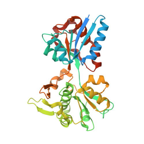
Apo and ligand-bound structures of ModA from the archaeon Methanosarcina acetivorans

Chan, S.Giuroiu, I.Chernishof, I.Sawaya, M.R.Chiang, J.Gunsalus, R.P.Arbing, M.A.Perry, L.J.

(2010) Acta Crystallogr Sect F Struct Biol Cryst Commun 66: 242-250

  • DOI: https://doi.org/10.1107/S1744309109055158
  • Primary Citation of Related Structures:  
    3K6U, 3K6V, 3K6W, 3K6X

  • PubMed Abstract: 

    The trace-element oxyanion molybdate, which is required for the growth of many bacterial and archaeal species, is transported into the cell by an ATP-binding cassette (ABC) transporter superfamily uptake system called ModABC. ModABC consists of the ModA periplasmic solute-binding protein, the integral membrane-transport protein ModB and the ATP-binding and hydrolysis cassette protein ModC. In this study, X-ray crystal structures of ModA from the archaeon Methanosarcina acetivorans (MaModA) have been determined in the apoprotein conformation at 1.95 and 1.69 A resolution and in the molybdate-bound conformation at 2.25 and 2.45 A resolution. The overall domain structure of MaModA is similar to other ModA proteins in that it has a bilobal structure in which two mixed alpha/beta domains are linked by a hinge region. The apo MaModA is the first unliganded archaeal ModA structure to be determined: it exhibits a deep cleft between the two domains and confirms that upon binding ligand one domain is rotated towards the other by a hinge-bending motion, which is consistent with the 'Venus flytrap' model seen for bacterial-type periplasmic binding proteins. In contrast to the bacterial ModA structures, which have tetrahedral coordination of their metal substrates, molybdate-bound MaModA employs octahedral coordination of its substrate like other archaeal ModA proteins.


  • Organizational Affiliation

    UCLA-DOE Institute for Genomics and Proteomics, University of California at Los Angeles, Los Angeles, CA 90095, USA.


Macromolecules
Find similar proteins by:  (by identity cutoff)  |  3D Structure
Entity ID: 1
MoleculeChains Sequence LengthOrganismDetailsImage
Solute-binding protein MA_0280354Methanosarcina acetivoransMutation(s): 0 
Gene Names: MA0280MA_0280
UniProt
Find proteins for Q8TTZ5 (Methanosarcina acetivorans (strain ATCC 35395 / DSM 2834 / JCM 12185 / C2A))
Explore Q8TTZ5 
Go to UniProtKB:  Q8TTZ5
Entity Groups  
Sequence Clusters30% Identity50% Identity70% Identity90% Identity95% Identity100% Identity
UniProt GroupQ8TTZ5
Sequence Annotations
Expand
  • Reference Sequence
Small Molecules
Ligands 1 Unique
IDChains Name / Formula / InChI Key2D Diagram3D Interactions
CIT
Query on CIT

Download Ideal Coordinates CCD File 
B [auth A]CITRIC ACID
C6 H8 O7
KRKNYBCHXYNGOX-UHFFFAOYSA-N
Experimental Data & Validation

Experimental Data

  • Method: X-RAY DIFFRACTION
  • Resolution: 1.69 Å
  • R-Value Free: 0.211 
  • R-Value Work: 0.189 
  • R-Value Observed: 0.190 
  • Space Group: P 61
Unit Cell:
Length ( Å )Angle ( ˚ )
a = 81.629α = 90
b = 81.629β = 90
c = 104.636γ = 120
Software Package:
Software NamePurpose
DENZOdata reduction
SCALEPACKdata scaling
EPMRphasing
REFMACrefinement
PDB_EXTRACTdata extraction
CrystalCleardata collection

Structure Validation

View Full Validation Report


Entry History 

Deposition Data

Revision History  (Full details and data files)

  • Version 1.0: 2010-01-12
    Type: Initial release
  • Version 1.1: 2011-07-13
    Changes: Advisory, Version format compliance
  • Version 1.2: 2017-11-01
    Changes: Advisory, Refinement description
  • Version 1.3: 2024-02-21
    Changes: Advisory, Data collection, Database references, Derived calculations