


The structure of E.coli peptide deformylase (PDF) in complex with peptidomimetic ligand BB2827
Cheng, R.K.Y., Crawley, L., Wood, M., Barker, J., Felicetti, B., Whittaker, M.To be published.
Experimental Data Snapshot
Entity ID: 1 | |||||
|---|---|---|---|---|---|
| Molecule | Chains | Sequence Length | Organism | Details | Image |
| Peptide deformylase | 169 | Escherichia coli K-12 | Mutation(s): 0 Gene Names: def, fms, b3287, JW3248 EC: 3.5.1.88 | 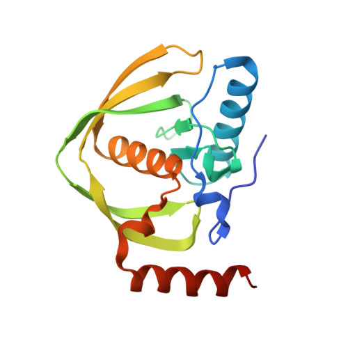 | |
UniProt | |||||
Find proteins for P0A6K3 (Escherichia coli (strain K12)) Explore P0A6K3 Go to UniProtKB: P0A6K3 | |||||
Entity Groups | |||||
| Sequence Clusters | 30% Identity50% Identity70% Identity90% Identity95% Identity100% Identity | ||||
| UniProt Group | P0A6K3 | ||||
Sequence AnnotationsExpand | |||||
| |||||
| Ligands 2 Unique | |||||
|---|---|---|---|---|---|
| ID | Chains | Name / Formula / InChI Key | 2D Diagram | 3D Interactions | |
| 2BB Query on 2BB | E [auth A], G [auth B] | (2S,3R)-N~4~-[(1S)-1-(dimethylcarbamoyl)-2,2-dimethylpropyl]-N~1~,2-dihydroxy-3-(2-methylpropyl)butanediamide C16 H31 N3 O5 USHCFFHZEHRVBD-GRYCIOLGSA-N |  | ||
| NI Query on NI | D [auth A], F [auth B], H [auth C] | NICKEL (II) ION Ni VEQPNABPJHWNSG-UHFFFAOYSA-N |  | ||
| Length ( Å ) | Angle ( ˚ ) |
|---|---|
| a = 73.96 | α = 90 |
| b = 73.96 | β = 90 |
| c = 241.5 | γ = 90 |
| Software Name | Purpose |
|---|---|
| DNA | data collection |
| PHASER | phasing |
| REFMAC | refinement |
| MOSFLM | data reduction |
| SCALA | data scaling |
RCSB PDB (citation) is hosted by
RCSB PDB is a member of the

