3K5D

Crystal Structure of BACE-1 in complex with AHM178


Experimental Data Snapshot

  • Method: X-RAY DIFFRACTION
  • Resolution: 2.90 Å
  • R-Value Free: 0.238 
  • R-Value Work: 0.202 
  • R-Value Observed: 0.204 

wwPDB Validation   3D Report Full Report


Ligand Structure Quality Assessment 


This is version 1.3 of the entry. See complete history


Literature

Structure-based design and synthesis of novel P2/P3 modified, non-peptidic beta-secretase (BACE-1) inhibitors.

Hanessian, S.Shao, Z.Betschart, C.Rondeau, J.M.Neumann, U.Tintelnot-Blomley, M.

(2010) Bioorg Med Chem Lett 20: 1924-1927

  • DOI: https://doi.org/10.1016/j.bmcl.2010.01.139
  • Primary Citation of Related Structures:  
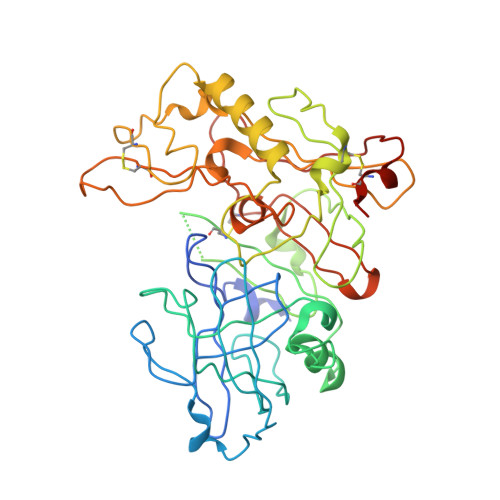
    3K5D, 3K5F, 3K5G

  • PubMed Abstract: 

    Starting from peptidomimetic BACE-1 inhibitors, the P2 amino acid including the P2/P3 peptide bond was replaced by a rigid 3-aminomethyl cyclohexane carboxylic acid. Co-crystallization revealed an unexpected binding mode with the P3/P4 amide bond placed into the S3 pocket resulting in a new hydrogen bond interaction pattern. Further optimization based on this structure resulted in highly potent BACE-1 inhibitors with selectivity over BACE-2 and cathepsin D.


  • Organizational Affiliation

    Department of Chemistry, Université de Montréal, Montréal, Canada. stephen.hanessian@umontreal.ca


Macromolecules
Find similar proteins by:  (by identity cutoff)  |  3D Structure
Entity ID: 1
MoleculeChains Sequence LengthOrganismDetailsImage
Beta-secretase 1
A, B, C
408Homo sapiensMutation(s): 0 
Gene Names: BACEBACE1KIAA1149
EC: 3.4.23.46
UniProt & NIH Common Fund Data Resources
Find proteins for P56817 (Homo sapiens)
Explore P56817 
Go to UniProtKB:  P56817
PHAROS:  P56817
GTEx:  ENSG00000186318 
Entity Groups  
Sequence Clusters30% Identity50% Identity70% Identity90% Identity95% Identity100% Identity
UniProt GroupP56817
Sequence Annotations
Expand
  • Reference Sequence
Small Molecules
Ligands 1 Unique
IDChains Name / Formula / InChI Key2D Diagram3D Interactions
XLI
Query on XLI

Download Ideal Coordinates CCD File 
D [auth A],
E [auth B],
F [auth C]
N-acetyl-L-leucyl-N-[(4S,5S,7R)-8-(butylamino)-5-hydroxy-2,7-dimethyl-8-oxooctan-4-yl]-L-methioninamide
C27 H52 N4 O5 S
PEWRQHFGBBTOMS-FFYZIMEISA-N
Binding Affinity Annotations 
IDSourceBinding Affinity
XLI PDBBind:  3K5D IC50: 53 (nM) from 1 assay(s)
BindingDB:  3K5D IC50: 53 (nM) from 1 assay(s)
Biologically Interesting Molecules (External Reference) 1 Unique
Experimental Data & Validation

Experimental Data

  • Method: X-RAY DIFFRACTION
  • Resolution: 2.90 Å
  • R-Value Free: 0.238 
  • R-Value Work: 0.202 
  • R-Value Observed: 0.204 
  • Space Group: P 1 21 1
Unit Cell:
Length ( Å )Angle ( ˚ )
a = 82.547α = 90
b = 101.299β = 104.16
c = 102.218γ = 90
Software Package:
Software NamePurpose
CNXrefinement
DENZOdata reduction
SCALEPACKdata scaling
CNXphasing

Structure Validation

View Full Validation Report


Ligand Structure Quality Assessment 


Entry History 

Deposition Data

Revision History  (Full details and data files)

  • Version 1.0: 2010-05-05
    Type: Initial release
  • Version 1.1: 2011-07-13
    Changes: Atomic model, Database references, Derived calculations, Non-polymer description, Structure summary, Version format compliance
  • Version 1.2: 2012-12-12
    Changes: Other
  • Version 1.3: 2017-11-01
    Changes: Refinement description