3K3P

Crystal Structure of the Apo Form of D-Alanine:D-Alanine Ligase (DDl) from Streptococcus mutans


Experimental Data Snapshot

  • Method: X-RAY DIFFRACTION
  • Resolution: 2.23 Å
  • R-Value Free: 0.248 
  • R-Value Work: 0.196 
  • R-Value Observed: 0.199 

wwPDB Validation   3D Report Full Report


This is version 1.3 of the entry. See complete history


Literature

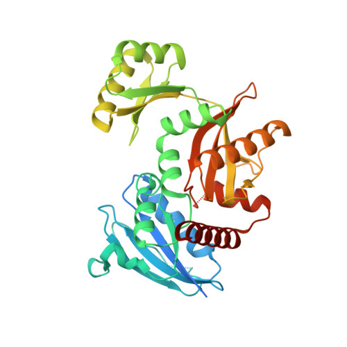
Crystal structure of the Apo form of D-Alanine:D-Alanine ligase (DDl) from Streptococcus mutans.

Lu, Y.Xu, H.Zhao, X.

(2010) Protein Pept Lett 17: 1053-1057

  • DOI: https://doi.org/10.2174/092986610791498858
  • Primary Citation of Related Structures:  
    3K3P

  • PubMed Abstract: 

    D-Alanine:D-Alanine ligase (DDl) catalyzes the formation of D-Alanine:D-Alanine dipeptide and is an essential enzyme in bacterial cell wall biosynthesis.. This enzyme does not have a human ortholog, making it an attractive target for developing new antibiotic drugs. We determined the crystal structure at 2.23 A resolution of DDl from Streptococcus mutans (SmDDl), the principal aetiological agent of human dental caries. This structure reveals that SmDDl is a dimer and has a disordered omega-loop region.


  • Organizational Affiliation

    West China Hospital Nanomedicine Laboratory, Center for Regenerative Medicine and Institute for Nanobiomedical Technology and Membrane Biology, West China Hospital Sichuan University, Chengdu 610065, Sichuan, China.


Macromolecules
Find similar proteins by:  (by identity cutoff)  |  3D Structure
Entity ID: 1
MoleculeChains Sequence LengthOrganismDetailsImage
D-alanine--D-alanine ligase
A, B
383Streptococcus mutansMutation(s): 0 
Gene Names: ddl
EC: 6.3.2.4
UniProt
Find proteins for P95803 (Streptococcus mutans serotype c (strain ATCC 700610 / UA159))
Explore P95803 
Go to UniProtKB:  P95803
Entity Groups  
Sequence Clusters30% Identity50% Identity70% Identity90% Identity95% Identity100% Identity
UniProt GroupP95803
Sequence Annotations
Expand
  • Reference Sequence
Experimental Data & Validation

Experimental Data

  • Method: X-RAY DIFFRACTION
  • Resolution: 2.23 Å
  • R-Value Free: 0.248 
  • R-Value Work: 0.196 
  • R-Value Observed: 0.199 
  • Space Group: P 32
Unit Cell:
Length ( Å )Angle ( ˚ )
a = 79.502α = 90
b = 79.502β = 90
c = 109.4γ = 120
Software Package:
Software NamePurpose
REFMACrefinement
PDB_EXTRACTdata extraction
CrysalisProdata reduction
SCALAdata scaling
MOLREPphasing

Structure Validation

View Full Validation Report


Entry History 

Deposition Data

  • Released Date: 2010-09-15 
  • Deposition Author(s): Lu, Y.

Revision History  (Full details and data files)

  • Version 1.0: 2010-09-15
    Type: Initial release
  • Version 1.1: 2011-07-13
    Changes: Version format compliance
  • Version 1.2: 2019-10-16
    Changes: Advisory, Data collection, Database references
  • Version 1.3: 2023-11-01
    Changes: Advisory, Data collection, Database references, Refinement description