3K3K

Crystal structure of dimeric abscisic acid (ABA) receptor pyrabactin resistance 1 (PYR1) with ABA-bound closed-lid and ABA-free open-lid subunits


Experimental Data Snapshot

  • Method: X-RAY DIFFRACTION
  • Resolution: 1.70 Å
  • R-Value Free: 0.237 
  • R-Value Work: 0.189 
  • R-Value Observed: 0.191 

wwPDB Validation   3D Report Full Report


Ligand Structure Quality Assessment 


This is version 1.2 of the entry. See complete history


Literature

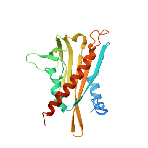
Structural mechanism of abscisic acid binding and signaling by dimeric PYR1.

Nishimura, N.Hitomi, K.Arvai, A.S.Rambo, R.P.Hitomi, C.Cutler, S.R.Schroeder, J.I.Getzoff, E.D.

(2009) Science 326: 1373-1379

  • DOI: https://doi.org/10.1126/science.1181829
  • Primary Citation of Related Structures:  
    3K3K

  • PubMed Abstract: 

    The phytohormone abscisic acid (ABA) acts in seed dormancy, plant development, drought tolerance, and adaptive responses to environmental stresses. Structural mechanisms mediating ABA receptor recognition and signaling remain unknown but are essential for understanding and manipulating abiotic stress resistance. Here, we report structures of pyrabactin resistance 1 (PYR1), a prototypical PYR/PYR1-like (PYL)/regulatory component of ABA receptor (RCAR) protein that functions in early ABA signaling. The crystallographic structure reveals an alpha/beta helix-grip fold and homodimeric assembly, verified in vivo by coimmunoprecipitation. ABA binding within a large internal cavity switches structural motifs distinguishing ABA-free "open-lid" from ABA-bound "closed-lid" conformations. Small-angle x-ray scattering suggests that ABA signals by converting PYR1 to a more compact, symmetric closed-lid dimer. Site-directed PYR1 mutants designed to disrupt hormone binding lose ABA-triggered interactions with type 2C protein phosphatase partners in planta.


  • Organizational Affiliation

    Division of Biological Sciences, Cell and Developmental Biology Section, University of California at San Diego, La Jolla, CA 92093, USA.


Macromolecules
Find similar proteins by:  (by identity cutoff)  |  3D Structure
Entity ID: 1
MoleculeChains Sequence LengthOrganismDetailsImage
Abscisic acid receptor PYR1
A, B
211Arabidopsis thalianaMutation(s): 0 
Gene Names: AT4g17870PYR1T6K21.50
UniProt
Find proteins for O49686 (Arabidopsis thaliana)
Explore O49686 
Go to UniProtKB:  O49686
Entity Groups  
Sequence Clusters30% Identity50% Identity70% Identity90% Identity95% Identity100% Identity
UniProt GroupO49686
Sequence Annotations
Expand
  • Reference Sequence
Small Molecules
Ligands 1 Unique
IDChains Name / Formula / InChI Key2D Diagram3D Interactions
A8S
Query on A8S

Download Ideal Coordinates CCD File 
C [auth B](2Z,4E)-5-[(1S)-1-hydroxy-2,6,6-trimethyl-4-oxocyclohex-2-en-1-yl]-3-methylpenta-2,4-dienoic acid
C15 H20 O4
JLIDBLDQVAYHNE-YKALOCIXSA-N
Experimental Data & Validation

Experimental Data

  • Method: X-RAY DIFFRACTION
  • Resolution: 1.70 Å
  • R-Value Free: 0.237 
  • R-Value Work: 0.189 
  • R-Value Observed: 0.191 
  • Space Group: P 1 2 1
Unit Cell:
Length ( Å )Angle ( ˚ )
a = 50.208α = 90
b = 61.407β = 98.97
c = 82.844γ = 90
Software Package:
Software NamePurpose
REFMACrefinement
PDB_EXTRACTdata extraction
HKL-2000data collection
HKL-2000data reduction
HKL-2000data scaling
PHASERphasing

Structure Validation

View Full Validation Report


Ligand Structure Quality Assessment 


Entry History 

Deposition Data

Revision History  (Full details and data files)

  • Version 1.0: 2009-11-17
    Type: Initial release
  • Version 1.1: 2011-07-13
    Changes: Non-polymer description, Version format compliance
  • Version 1.2: 2023-09-06
    Changes: Data collection, Database references, Derived calculations, Refinement description, Structure summary