3K3E

Crystal structure of the PDE9A catalytic domain in complex with (R)-BAY73-6691


Experimental Data Snapshot

  • Method: X-RAY DIFFRACTION
  • Resolution: 2.70 Å
  • R-Value Free: 0.258 
  • R-Value Work: 0.230 
  • R-Value Observed: 0.230 

wwPDB Validation   3D Report Full Report


Ligand Structure Quality Assessment 


This is version 1.2 of the entry. See complete history


Literature

Insight into Binding of Phosphodiesterase-9A Selective Inhibitors by Crystal Structures and Mutagenesis

Wang, H.Luo, X.Ye, M.Hou, J.Robinson, H.Ke, H.

(2010) J Med Chem 53: 1726-1731

  • DOI: https://doi.org/10.1021/jm901519f
  • Primary Citation of Related Structures:  
    3K3E, 3K3H

  • PubMed Abstract: 

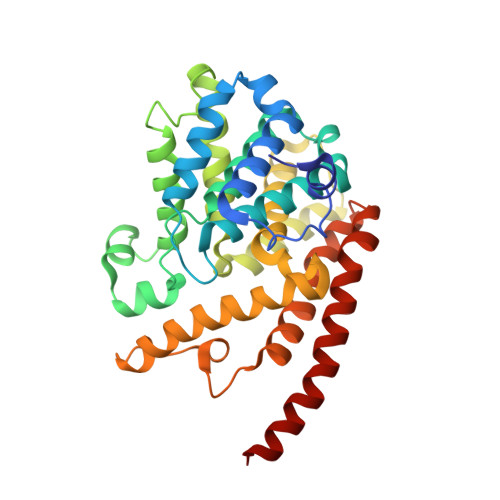
    PDE9 inhibitors have been studied as therapeutics for treatment of cardiovascular diseases, diabetes, and neurodegenerative disorders. To illustrate the inhibitor selectivity, the crystal structures of the PDE9A catalytic domain in complex with the enantiomers of PDE9 inhibitor 1-(2-chlorophenyl)-6-(3,3,3-trifluoro-2-methylpropyl)-1H-pyrazolo[3,4-d]pyrimidine-4(5H)-one ((R)-BAY73-6691 or (S)-BAY73-6691, 1r or 1s) were determined and mutagenesis was performed. The structures showed that the fluoromethyl groups of 1r and 1s had different orientations while the other parts of the inhibitors commonly interacted with PDE9A. These differences may explain the slightly different affinity of 1r (IC(50) = 22 nM) and 1s (IC(50) = 88 nM). The mutagenesis experiments revealed that contribution of the binding residues to the inhibitor sensitivity varies dramatically, from few-fold to 3 orders of magnitude. On the basis of the crystal structures, a hypothesized compound that simulates the recently published PDE9 inhibitors was modeled to provide insight into the inhibitor selectivity.


  • Organizational Affiliation

    Department of Biochemistry and Biophysics and Lineberger Comprehensive Cancer Center, The University of North Carolina, Chapel Hill, North Carolina 27599-7260, USA.


Macromolecules
Find similar proteins by:  (by identity cutoff)  |  3D Structure
Entity ID: 1
MoleculeChains Sequence LengthOrganismDetailsImage
High affinity cGMP-specific 3',5'-cyclic phosphodiesterase 9A
A, B
326Homo sapiensMutation(s): 0 
Gene Names: PDE9APDE9A2
EC: 3.1.4.35
UniProt & NIH Common Fund Data Resources
Find proteins for O76083 (Homo sapiens)
Explore O76083 
Go to UniProtKB:  O76083
PHAROS:  O76083
GTEx:  ENSG00000160191 
Entity Groups  
Sequence Clusters30% Identity50% Identity70% Identity90% Identity95% Identity100% Identity
UniProt GroupO76083
Sequence Annotations
Expand
  • Reference Sequence
Experimental Data & Validation

Experimental Data

  • Method: X-RAY DIFFRACTION
  • Resolution: 2.70 Å
  • R-Value Free: 0.258 
  • R-Value Work: 0.230 
  • R-Value Observed: 0.230 
  • Space Group: P 41 21 2
Unit Cell:
Length ( Å )Angle ( ˚ )
a = 103.19α = 90
b = 103.19β = 90
c = 270.849γ = 90
Software Package:
Software NamePurpose
ADSCdata collection
AMoREphasing
CNSrefinement
HKL-2000data reduction
HKL-2000data scaling

Structure Validation

View Full Validation Report


Ligand Structure Quality Assessment 


Entry History 

Deposition Data

Revision History  (Full details and data files)

  • Version 1.0: 2010-02-16
    Type: Initial release
  • Version 1.1: 2011-07-13
    Changes: Version format compliance
  • Version 1.2: 2013-11-13
    Changes: Non-polymer description