3K37

Crystal Structure of B/Perth Neuraminidase in complex with Peramivir


Experimental Data Snapshot

  • Method: X-RAY DIFFRACTION
  • Resolution: 2.00 Å
  • R-Value Free: 0.237 
  • R-Value Work: 0.171 
  • R-Value Observed: 0.175 

wwPDB Validation   3D Report Full Report


Ligand Structure Quality Assessment 


This is version 2.1 of the entry. See complete history


Literature

Structural and Functional Basis of Resistance to Neuraminidase Inhibitors of Influenza B Viruses.

Oakley, A.J.Barrett, S.Peat, T.S.Newman, J.Streltsov, V.A.Waddington, L.Saito, T.Tashiro, M.McKimm-Breschkin, J.L.

(2010) J Med Chem 

  • DOI: https://doi.org/10.1021/jm100621s
  • Primary Citation of Related Structures:  
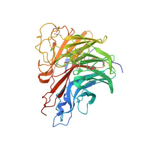
    3K36, 3K37, 3K38, 3K39, 3K3A

  • PubMed Abstract: 

    We have identified a virus, B/Perth/211/2001, with a spontaneous mutation, D197E in the neuraminidase (NA), which confers cross-resistance to all NA inhibitors. We analyzed enzyme properties of the D197 and E197 NAs and compared these to a D197N NA, known to arise after oseltamivir treatment. Zanamivir and peramivir bound slowly to the wild type NA, but binding of oseltamivir was more rapid. The D197E/N mutations resulted in faster binding of all three inhibitors. Analysis of the crystal structures of D197 and E197 NAs with and without inhibitors showed that the D197E mutation compromised the interaction of neighboring R150 with the N-acetyl group, common to the substrate sialic acid and all NA inhibitors. Although rotation of the E275 in the NA active site occurs upon binding peramivir in both the D197 and E197 NAs, this does not occur upon binding oseltamivir in the E197 NA. Lack of the E275 rotation would also account for the loss of slow binding and the partial resistance of influenza B wild type NAs to oseltamivir.


  • Organizational Affiliation

    CSIRO Materials Science and Engineering, Parkville, 343 Royal Parade, Parkville, Victoria, 3052, Australia.


Macromolecules
Find similar proteins by:  (by identity cutoff)  |  3D Structure
Entity ID: 1
MoleculeChains Sequence LengthOrganismDetailsImage
Neuraminidase
A, B
397Influenza B virus (B/Perth/211/2001)Mutation(s): 0 
EC: 3.2.1.18
UniProt
Find proteins for Q3S340 (Influenza B virus)
Explore Q3S340 
Go to UniProtKB:  Q3S340
Entity Groups  
Sequence Clusters30% Identity50% Identity70% Identity90% Identity95% Identity100% Identity
UniProt GroupQ3S340
Sequence Annotations
Expand
  • Reference Sequence
Oligosaccharides

Help

Entity ID: 2
MoleculeChains Length2D Diagram Glycosylation3D Interactions
2-acetamido-2-deoxy-beta-D-glucopyranose-(1-4)-2-acetamido-2-deoxy-beta-D-glucopyranose
C, D
2N-Glycosylation
Glycosylation Resources
GlyTouCan:  G42666HT
GlyCosmos:  G42666HT
GlyGen:  G42666HT
Small Molecules
Binding Affinity Annotations 
IDSourceBinding Affinity
BCZ PDBBind:  3K37 Ki: 1.2 (nM) from 1 assay(s)
Binding MOAD:  3K37 Ki: 1.2 (nM) from 1 assay(s)
Experimental Data & Validation

Experimental Data

  • Method: X-RAY DIFFRACTION
  • Resolution: 2.00 Å
  • R-Value Free: 0.237 
  • R-Value Work: 0.171 
  • R-Value Observed: 0.175 
  • Space Group: I 4
Unit Cell:
Length ( Å )Angle ( ˚ )
a = 88.645α = 90
b = 88.645β = 90
c = 207.523γ = 90
Software Package:
Software NamePurpose
Blu-Icedata collection
MOLREPphasing
REFMACrefinement
MOSFLMdata reduction
SCALAdata scaling

Structure Validation

View Full Validation Report


Ligand Structure Quality Assessment 


Entry History 

Deposition Data

Revision History  (Full details and data files)

  • Version 1.0: 2010-09-01
    Type: Initial release
  • Version 1.1: 2011-07-13
    Changes: Version format compliance
  • Version 2.0: 2020-07-29
    Type: Remediation
    Reason: Carbohydrate remediation
    Changes: Atomic model, Data collection, Derived calculations, Structure summary
  • Version 2.1: 2023-11-01
    Changes: Data collection, Database references, Refinement description, Structure summary