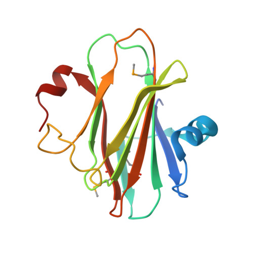
Crystal structure and oligomeric state of the RetS signaling kinase sensory domain.
Jing, X., Jaw, J., Robinson, H.H., Schubot, F.D.(2010) Proteins 78: 1631-1640
- PubMed: 20112417
- DOI: https://doi.org/10.1002/prot.22679
- Primary Citation of Related Structures:
3JYB - PubMed Abstract:
The opportunistic pathogen Pseudomonas aeruginosa may cause both acute and chronic-persistent infections in predisposed individuals. Acute infections require the presence of a functional type III secretion system (T3SS), whereas chronic P. aeruginosa infections are characterized by the formation of drug-resistant biofilms. The T3SS and biofilm formation are reciprocally regulated by the signaling kinases LadS, RetS, and GacS. RetS downregulates biofilm formation and upregulates expression of the T3SS through a unique mechanism. RetS forms a heterodimeric complex with GacS and thus prevents GacS autophosphorylation and downstream signaling. The signals that regulate RetS are not known but RetS possesses a distinctive periplasmic sensor domain that is believed to serve as receptor for the regulatory ligand. We have determined the crystal structure of the RetS sensory domain at 2.0 A resolution. The structure closely resembles those of carbohydrate binding modules of other proteins, suggesting that the elusive ligands are likely carbohydrate moieties. In addition to the conserved beta-sandwich structure, the sensory domain features two alpha helices which create a unique surface topology. Protein-protein crosslinking and fluorescence energy transfer experiments also revealed that the sensory domain dimerizes with a dissociation constant of K(d) = 580 +/- 50 nM, a result with interesting implications for our understanding of the underlying signaling mechanism.
Organizational Affiliation:
Department of Biological Sciences, Life Science I, Virginia Polytechnic Institute and State University, Blacksburg, Virginia 24060, USA.