3JWQ

Crystal structure of chimeric PDE5/PDE6 catalytic domain complexed with sildenafil


Experimental Data Snapshot

  • Method: X-RAY DIFFRACTION
  • Resolution: 2.87 Å
  • R-Value Free: 0.275 
  • R-Value Work: 0.223 
  • R-Value Observed: 0.226 

wwPDB Validation   3D Report Full Report


Ligand Structure Quality Assessment 


This is version 1.3 of the entry. See complete history


Literature

Structural basis of phosphodiesterase 6 inhibition by the C-terminal region of the gamma-subunit

Barren, B.Gakhar, L.Muradov, H.Boyd, K.K.Ramaswamy, S.Artemyev, N.O.

(2009) EMBO J 28: 3613-3622

  • DOI: https://doi.org/10.1038/emboj.2009.284
  • Primary Citation of Related Structures:  
    3JWQ, 3JWR

  • PubMed Abstract: 

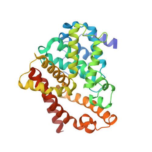
    The inhibitory interaction of phosphodiesterase-6 (PDE6) with its gamma-subunit (Pgamma) is pivotal in vertebrate phototransduction. Here, crystal structures of a chimaeric PDE5/PDE6 catalytic domain (PDE5/6cd) complexed with sildenafil or 3-isobutyl-1-methylxanthine and the Pgamma-inhibitory peptide Pgamma(70-87) have been determined at 2.9 and 3.0 A, respectively. These structures show the determinants and the mechanism of the PDE6 inhibition by Pgamma and suggest the conformational change of Pgamma on transducin activation. Two variable H- and M-loops of PDE5/6cd form a distinct interface that contributes to the Pgamma-binding site. This allows the Pgamma C-terminus to fit into the opening of the catalytic pocket, blocking cGMP access to the active site. Our analysis suggests that disruption of the H-M loop interface and Pgamma-binding site is a molecular cause of retinal degeneration in atrd3 mice. Comparison of the two PDE5/6cd structures shows an overlap between the sildenafil and Pgamma(70-87)-binding sites, thereby providing critical insights into the side effects of PDE5 inhibitors on vision.


  • Organizational Affiliation

    Department of Molecular Physiology and Biophysics, University of Iowa, Iowa City, IA, USA.


Macromolecules
Find similar proteins by:  (by identity cutoff)  |  3D Structure
Entity ID: 1
MoleculeChains Sequence LengthOrganismDetailsImage
cGMP-specific 3',5'-cyclic phosphodiesterase catalytic domain, Cone cGMP-specific 3',5'-cyclic phosphodiesterase subunit alpha chimera
A, B, C, D
330Homo sapiensMutation(s): 0 
Gene Names: PDE5APDE5PDE6CPDEA2
EC: 3.1.4.35
UniProt & NIH Common Fund Data Resources
Find proteins for O76074 (Homo sapiens)
Explore O76074 
Go to UniProtKB:  O76074
PHAROS:  O76074
GTEx:  ENSG00000138735 
Find proteins for P51160 (Homo sapiens)
Explore P51160 
Go to UniProtKB:  P51160
PHAROS:  P51160
GTEx:  ENSG00000095464 
Entity Groups  
Sequence Clusters30% Identity50% Identity70% Identity90% Identity95% Identity100% Identity
UniProt GroupsP51160O76074
Sequence Annotations
Expand
  • Reference Sequence
Small Molecules
Ligands 3 Unique
IDChains Name / Formula / InChI Key2D Diagram3D Interactions
VIA
Query on VIA

Download Ideal Coordinates CCD File 
G [auth A],
J [auth B],
M [auth C],
P [auth D]
5-{2-ETHOXY-5-[(4-METHYLPIPERAZIN-1-YL)SULFONYL]PHENYL}-1-METHYL-3-PROPYL-1H,6H,7H-PYRAZOLO[4,3-D]PYRIMIDIN-7-ONE
C22 H30 N6 O4 S
BNRNXUUZRGQAQC-UHFFFAOYSA-N
ZN
Query on ZN

Download Ideal Coordinates CCD File 
E [auth A],
H [auth B],
K [auth C],
N [auth D]
ZINC ION
Zn
PTFCDOFLOPIGGS-UHFFFAOYSA-N
MG
Query on MG

Download Ideal Coordinates CCD File 
F [auth A],
I [auth B],
L [auth C],
O [auth D]
MAGNESIUM ION
Mg
JLVVSXFLKOJNIY-UHFFFAOYSA-N
Binding Affinity Annotations 
IDSourceBinding Affinity
VIA BindingDB:  3JWQ Ki: min: 1.6, max: 10 (nM) from 4 assay(s)
IC50: min: 0.3, max: 1000 (nM) from 31 assay(s)
EC50: min: 8.7, max: 1000 (nM) from 3 assay(s)
PDBBind:  3JWQ Ki: 25 (nM) from 1 assay(s)
Experimental Data & Validation

Experimental Data

  • Method: X-RAY DIFFRACTION
  • Resolution: 2.87 Å
  • R-Value Free: 0.275 
  • R-Value Work: 0.223 
  • R-Value Observed: 0.226 
  • Space Group: P 21 21 21
Unit Cell:
Length ( Å )Angle ( ˚ )
a = 75.732α = 90
b = 109.825β = 90
c = 199.668γ = 90
Software Package:
Software NamePurpose
ADSCdata collection
PHASERphasing
REFMACrefinement
d*TREKdata reduction
d*TREKdata scaling

Structure Validation

View Full Validation Report


Ligand Structure Quality Assessment 


Entry History 

Deposition Data

Revision History  (Full details and data files)

  • Version 1.0: 2009-10-13
    Type: Initial release
  • Version 1.1: 2011-07-13
    Changes: Advisory, Version format compliance
  • Version 1.2: 2017-08-16
    Changes: Refinement description, Source and taxonomy
  • Version 1.3: 2023-09-06
    Changes: Data collection, Database references, Derived calculations, Refinement description, Structure summary