3JUB

Human gamma-glutamylamine cyclotransferase


Experimental Data Snapshot

  • Method: X-RAY DIFFRACTION
  • Resolution: 1.20 Å
  • R-Value Free: 0.149 
  • R-Value Work: 0.123 
  • R-Value Observed: 0.124 

wwPDB Validation   3D Report Full Report


This is version 1.2 of the entry. See complete history


Literature

Identification and characterization of {gamma}-glutamylamine cyclotransferase: An enzyme responsible for {gamma}-glutamyl-{epsilon}-lysine catabolism

Oakley, A.J.Coggan, M.Board, P.G.

(2010) J Biol Chem 285: 9642-9648

  • DOI: https://doi.org/10.1074/jbc.M109.082099
  • Primary Citation of Related Structures:  
    3JUB, 3JUC, 3JUD

  • PubMed Abstract: 

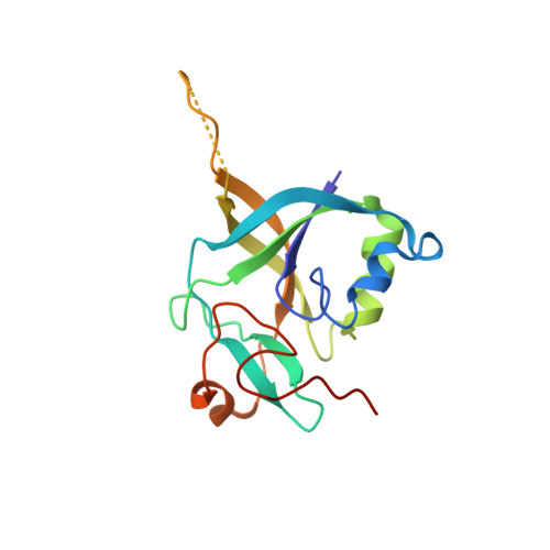
    Gamma-glutamylamine cyclotransferase (GGACT) is an enzyme that converts gamma-glutamylamines to free amines and 5-oxoproline. GGACT shows high activity toward gamma-glutamyl-epsilon-lysine, derived from the breakdown of fibrin and other proteins cross-linked by transglutaminases. The enzyme adopts the newly identified cyclotransferase fold, observed in gamma-glutamylcyclotransferase (GGCT), an enzyme with activity toward gamma-glutamyl-alpha-amino acids (Oakley, A. J., Yamada, T., Liu, D., Coggan, M., Clark, A. G., and Board, P. G. (2008) J. Biol. Chem. 283, 22031-22042). Despite the absence of significant sequence identity, several residues are conserved in the active sites of GGCT and GGACT, including a putative catalytic acid/base residue (GGACT Glu(82)). The structure of GGACT in complex with the reaction product 5-oxoproline provides evidence for a common catalytic mechanism in both enzymes. The proposed mechanism, combined with the three-dimensional structures, also explains the different substrate specificities of these enzymes. Despite significant sequence divergence, there are at least three subfamilies in prokaryotes and eukaryotes that have conserved the GGCT fold and GGCT enzymatic activity.


  • Organizational Affiliation

    Division of Molecular and Health Technologies, Commonwealth Scientific and Industrial Research Organization, Parkville, Victoria 3052.


Macromolecules
Find similar proteins by:  (by identity cutoff)  |  3D Structure
Entity ID: 1
MoleculeChains Sequence LengthOrganismDetailsImage
AIG2-like domain-containing protein 1153Homo sapiensMutation(s): 0 
Gene Names: A2LD1GGACT
EC: 2.3.2.4
UniProt & NIH Common Fund Data Resources
Find proteins for Q9BVM4 (Homo sapiens)
Explore Q9BVM4 
Go to UniProtKB:  Q9BVM4
PHAROS:  Q9BVM4
GTEx:  ENSG00000134864 
Entity Groups  
Sequence Clusters30% Identity50% Identity70% Identity90% Identity95% Identity100% Identity
UniProt GroupQ9BVM4
Sequence Annotations
Expand
  • Reference Sequence
Experimental Data & Validation

Experimental Data

  • Method: X-RAY DIFFRACTION
  • Resolution: 1.20 Å
  • R-Value Free: 0.149 
  • R-Value Work: 0.123 
  • R-Value Observed: 0.124 
  • Space Group: P 21 21 21
Unit Cell:
Length ( Å )Angle ( ˚ )
a = 35.995α = 90
b = 41.94β = 90
c = 84.012γ = 90
Software Package:
Software NamePurpose
Blu-Icedata collection
MOLREPphasing
REFMACrefinement
MOSFLMdata reduction
SCALAdata scaling

Structure Validation

View Full Validation Report


Entry History 

Deposition Data

  • Released Date: 2010-02-09 
  • Deposition Author(s): Oakley, A.J.

Revision History  (Full details and data files)

  • Version 1.0: 2010-02-09
    Type: Initial release
  • Version 1.1: 2011-07-13
    Changes: Version format compliance
  • Version 1.2: 2023-11-01
    Changes: Data collection, Database references, Derived calculations, Refinement description