


Crystal structure of Iron superoxide dismutase from Anaplasma phagocytophilum
Edwards, T.E., Staker, B.L., Seattle Structural Genomics Center for Infectious Disease (SSGCID)To be published.
Experimental Data Snapshot
wwPDB Validation 3D Report Full Report

Entity ID: 1 | |||||
|---|---|---|---|---|---|
| Molecule | Chains | Sequence Length | Organism | Details | Image |
| Superoxide dismutase | 227 | Anaplasma phagocytophilum str. HZ | Mutation(s): 0 Gene Names: sodB, APH_0371 EC: 1.15.1.1 | 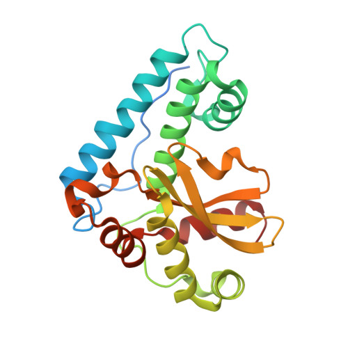 | |
UniProt | |||||
Find proteins for Q2GKX4 (Anaplasma phagocytophilum (strain HZ)) Explore Q2GKX4 Go to UniProtKB: Q2GKX4 | |||||
Entity Groups | |||||
| Sequence Clusters | 30% Identity50% Identity70% Identity90% Identity95% Identity100% Identity | ||||
| UniProt Group | Q2GKX4 | ||||
Sequence AnnotationsExpand | |||||
| |||||
| Ligands 2 Unique | |||||
|---|---|---|---|---|---|
| ID | Chains | Name / Formula / InChI Key | 2D Diagram | 3D Interactions | |
| FE Query on FE | E [auth A], G [auth B], I [auth C], K [auth D] | FE (III) ION Fe VTLYFUHAOXGGBS-UHFFFAOYSA-N |  | ||
| NA Query on NA | F [auth A], H [auth B], J [auth C], L [auth D] | SODIUM ION Na FKNQFGJONOIPTF-UHFFFAOYSA-N |  | ||
| Length ( Å ) | Angle ( ˚ ) |
|---|---|
| a = 45.66 | α = 102.09 |
| b = 66.6 | β = 104.83 |
| c = 85.44 | γ = 88.58 |
| Software Name | Purpose |
|---|---|
| XSCALE | data scaling |
| MOLREP | phasing |
| REFMAC | refinement |
| PDB_EXTRACT | data extraction |
| XDS | data reduction |
RCSB PDB (citation) is hosted by
RCSB PDB is a member of the

