3JRX

Crystal structure of the BC domain of ACC2 in complex with soraphen A


Experimental Data Snapshot

  • Method: X-RAY DIFFRACTION
  • Resolution: 2.50 Å
  • R-Value Free: 0.266 
  • R-Value Work: 0.221 
  • R-Value Observed: 0.221 

wwPDB Validation   3D Report Full Report


Ligand Structure Quality Assessment 


This is version 1.2 of the entry. See complete history


Literature

Molecular mechanism for the regulation of human ACC2 through phosphorylation by AMPK.

Cho, Y.S.Lee, J.I.Shin, D.Kim, H.T.Jung, H.Y.Lee, T.G.Kang, L.W.Ahn, Y.J.Cho, H.S.Heo, Y.S.

(2010) Biochem Biophys Res Commun 391: 187-192

  • DOI: https://doi.org/10.1016/j.bbrc.2009.11.029
  • Primary Citation of Related Structures:  
    3JRW, 3JRX

  • PubMed Abstract: 

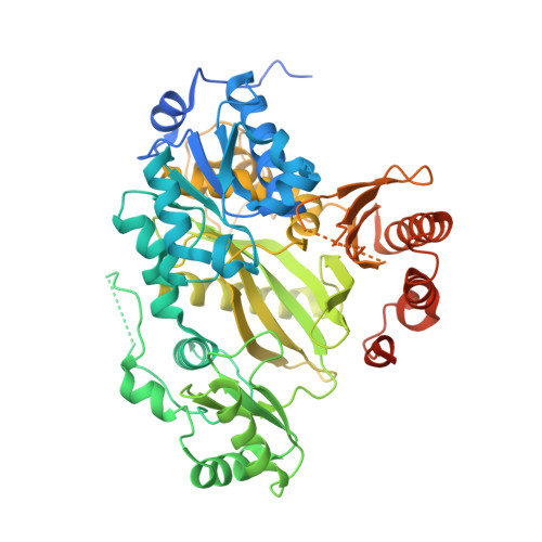
    Acetyl-CoA carboxylases (ACCs) have been highlighted as therapeutic targets for obesity and diabetes, as they play crucial roles in fatty acid metabolism. ACC activity is regulated through the short-term mechanism of inactivation by reversible phosphorylation. Here, we report the crystal structures of the biotin carboxylase (BC) domain of human ACC2 phosphorylated by AMP-activated protein kinase (AMPK). The phosphorylated Ser222 binds to the putative dimer interface of BC, disrupting polymerization and providing the molecular mechanism of inactivation by AMPK. We also determined the structure of the human BC domain in complex with soraphen A, a macrocyclic polyketide natural product. This structure shows that the compound binds to the binding site of phosphorylated Ser222, implying that its inhibition mechanism is the same as that of phosphorylation by AMPK.


  • Organizational Affiliation

    R&D Center, CrystalGenomics, Inc., Seoul 138-739, Republic of Korea.


Macromolecules
Find similar proteins by:  (by identity cutoff)  |  3D Structure
Entity ID: 1
MoleculeChains Sequence LengthOrganismDetailsImage
Acetyl-CoA carboxylase 2587Homo sapiensMutation(s): 0 
Gene Names: ACACBACC2ACCB
EC: 6.4.1.2 (PDB Primary Data), 6.3.4.14 (PDB Primary Data)
UniProt & NIH Common Fund Data Resources
Find proteins for O00763 (Homo sapiens)
Explore O00763 
Go to UniProtKB:  O00763
PHAROS:  O00763
GTEx:  ENSG00000076555 
Entity Groups  
Sequence Clusters30% Identity50% Identity70% Identity90% Identity95% Identity100% Identity
UniProt GroupO00763
Sequence Annotations
Expand
  • Reference Sequence
Small Molecules
Ligands 1 Unique
IDChains Name / Formula / InChI Key2D Diagram3D Interactions
S1A
Query on S1A

Download Ideal Coordinates CCD File 
B [auth A]SORAPHEN A
C29 H44 O8
WPMGNXPRKGXGBO-OFQQMTDKSA-N
Binding Affinity Annotations 
IDSourceBinding Affinity
S1A PDBBind:  3JRX Kd: 1 (nM) from 1 assay(s)
Binding MOAD:  3JRX Kd: 1 (nM) from 1 assay(s)
Experimental Data & Validation

Experimental Data

  • Method: X-RAY DIFFRACTION
  • Resolution: 2.50 Å
  • R-Value Free: 0.266 
  • R-Value Work: 0.221 
  • R-Value Observed: 0.221 
  • Space Group: P 32 2 1
Unit Cell:
Length ( Å )Angle ( ˚ )
a = 74.744α = 90
b = 74.744β = 90
c = 179.414γ = 120
Software Package:
Software NamePurpose
HKL-2000data collection
CNSrefinement
HKL-2000data reduction
HKL-2000data scaling
CNSphasing

Structure Validation

View Full Validation Report


Ligand Structure Quality Assessment 


Entry History 

Deposition Data

Revision History  (Full details and data files)

  • Version 1.0: 2010-01-12
    Type: Initial release
  • Version 1.1: 2011-07-13
    Changes: Version format compliance
  • Version 1.2: 2023-11-01
    Changes: Data collection, Database references, Derived calculations, Refinement description