3JQY

Crystal Structure of the polySia specific acetyltransferase NeuO


Experimental Data Snapshot

  • Method: X-RAY DIFFRACTION
  • Resolution: 1.70 Å
  • R-Value Free: 0.195 
  • R-Value Work: 0.166 
  • R-Value Observed: 0.167 

wwPDB Validation   3D Report Full Report


This is version 1.5 of the entry. See complete history


Literature

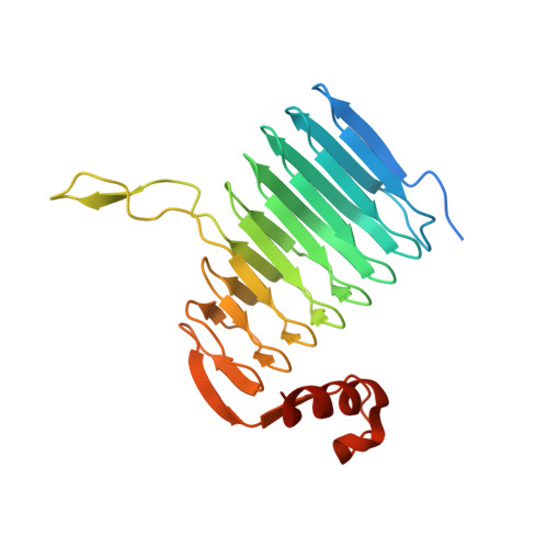
Crystal structure analysis of the polysialic acid specific O-acetyltransferase NeuO

Schulz, E.C.Bergfeld, A.K.Ficner, R.Muhlenhoff, M.

(2011) PLoS One 6: e17403-e17403

  • DOI: https://doi.org/10.1371/journal.pone.0017403
  • Primary Citation of Related Structures:  
    3JQY

  • PubMed Abstract: 

    The major virulence factor of the neuroinvasive pathogen Escherichia coli K1 is the K1 capsule composed of α2,8-linked polysialic acid (polySia). K1 strains harboring the CUS-3 prophage modify their capsular polysaccharide by phase-variable O-acetylation, a step that is associated with increased virulence. Here we present the crystal structure of the prophage-encoded polysialate O-acetyltransferase NeuO. The homotrimeric enzyme belongs to the left-handed β-helix (LβH) family of acyltransferases and is characterized by an unusual funnel-shaped outline. Comparison with other members of the LβH family allowed the identification of active site residues and proposal of a catalytic mechanism and highlighted structural characteristics of polySia specific O-acetyltransferases. As a unique feature of NeuO, the enzymatic activity linearly increases with the length of the N-terminal poly-ψ-domain which is composed of a variable number of tandem copies of an RLKTQDS heptad. Since the poly-ψ-domain was not resolved in the crystal structure it is assumed to be unfolded in the apo-enzyme.


  • Organizational Affiliation

    Abteilung für Molekulare Strukturbiologie, Institut für Mikrobiologie und Genetik, Georg-August-Universität Göttingen, Göttingen, Germany.


Macromolecules
Find similar proteins by:  (by identity cutoff)  |  3D Structure
Entity ID: 1
MoleculeChains Sequence LengthOrganismDetailsImage
Polysialic acid O-acetyltransferaseA [auth B],
B [auth A],
C
252Escherichia coliMutation(s): 0 
Gene Names: neuo
EC: 2.3.1.136
UniProt
Find proteins for A1ADJ6 (Escherichia coli O1:K1 / APEC)
Explore A1ADJ6 
Go to UniProtKB:  A1ADJ6
Entity Groups  
Sequence Clusters30% Identity50% Identity70% Identity90% Identity95% Identity100% Identity
UniProt GroupA1ADJ6
Sequence Annotations
Expand
  • Reference Sequence
Experimental Data & Validation

Experimental Data

  • Method: X-RAY DIFFRACTION
  • Resolution: 1.70 Å
  • R-Value Free: 0.195 
  • R-Value Work: 0.166 
  • R-Value Observed: 0.167 
  • Space Group: P 1 21 1
Unit Cell:
Length ( Å )Angle ( ˚ )
a = 62.833α = 90
b = 88.366β = 106.62
c = 72.996γ = 90
Software Package:
Software NamePurpose
DNAdata collection
MOLREPphasing
PHENIXrefinement
XDSdata reduction
SCALAdata scaling

Structure Validation

View Full Validation Report


Entry History 

Deposition Data

Revision History  (Full details and data files)

  • Version 1.0: 2010-08-18
    Type: Initial release
  • Version 1.1: 2011-07-13
    Changes: Version format compliance
  • Version 1.2: 2017-12-20
    Changes: Database references
  • Version 1.3: 2020-09-09
    Changes: Database references, Derived calculations, Structure summary
  • Version 1.4: 2024-03-20
    Changes: Data collection, Database references
  • Version 1.5: 2024-04-03
    Changes: Refinement description