3JQ4

The structure of the complex of the large ribosomal subunit from D. Radiodurans with the antibiotic lankacidin


Experimental Data Snapshot

  • Method: X-RAY DIFFRACTION
  • Resolution: 3.52 Å
  • R-Value Free: 0.325 
  • R-Value Work: 0.268 
  • R-Value Observed: 0.268 

wwPDB Validation   3D Report Full Report


This is version 1.3 of the entry. See complete history


Literature

The structure of ribosome-lankacidin complex reveals ribosomal sites for synergistic antibiotics

Auerbach, T.Mermershtain, I.Davidovich, C.Bashan, A.Belousoff, M.Wekselman, I.Zimmerman, E.Xiong, L.Klepacki, D.Arakawa, K.Kinashi, H.Mankin, A.S.Yonath, A.

(2010) Proc Natl Acad Sci U S A 107: 1983-1988

  • DOI: https://doi.org/10.1073/pnas.0914100107
  • Primary Citation of Related Structures:  
    3JQ4

  • PubMed Abstract: 

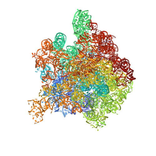
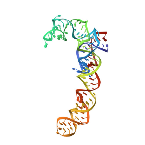
    Crystallographic analysis revealed that the 17-member polyketide antibiotic lankacidin produced by Streptomyces rochei binds at the peptidyl transferase center of the eubacterial large ribosomal subunit. Biochemical and functional studies verified this finding and showed interference with peptide bond formation. Chemical probing indicated that the macrolide lankamycin, a second antibiotic produced by the same species, binds at a neighboring site, at the ribosome exit tunnel. These two antibiotics can bind to the ribosome simultaneously and display synergy in inhibiting bacterial growth. The binding site of lankacidin and lankamycin partially overlap with the binding site of another pair of synergistic antibiotics, the streptogramins. Thus, at least two pairs of structurally dissimilar compounds have been selected in the course of evolution to act synergistically by targeting neighboring sites in the ribosome. These results underscore the importance of the corresponding ribosomal sites for development of clinically relevant synergistic antibiotics and demonstrate the utility of structural analysis for providing new directions for drug discovery.


  • Organizational Affiliation

    Department of Structural Biology, Weizmann Institute of Science, Rehovot 76100, Israel.


Macromolecules
Find similar nucleic acids by:  (by identity cutoff)  |  3D Structure
Entity ID: 1
MoleculeChains LengthOrganismImage
23S ribosomal RNA2,880Deinococcus radiodurans R1 = ATCC 13939 = DSM 20539
Sequence Annotations
Expand
  • Reference Sequence
Find similar nucleic acids by:  (by identity cutoff)  |  3D Structure
Entity ID: 2
MoleculeChains LengthOrganismImage
5S ribosomal RNA118Deinococcus radiodurans R1 = ATCC 13939 = DSM 20539
Sequence Annotations
Expand
  • Reference Sequence
Small Molecules
Ligands 1 Unique
IDChains Name / Formula / InChI Key2D Diagram3D Interactions
LC2
Query on LC2

Download Ideal Coordinates CCD File 
C [auth A]N-[(1S,2R,3E,5E,7S,9E,11E,13S,15R,19R)-7,13-dihydroxy-1,4,10,19-tetramethyl-17,18-dioxo-16-oxabicyclo[13.2.2]nonadeca-3,5,9,11-tetraen-2-yl]-2-oxopropanamide
C25 H33 N O7
ATDILMLBOZKFGI-JUTMVFGESA-N
Binding Affinity Annotations 
IDSourceBinding Affinity
LC2 PDBBind:  3JQ4 IC50: 1.00e+4 (nM) from 1 assay(s)
Experimental Data & Validation

Experimental Data

  • Method: X-RAY DIFFRACTION
  • Resolution: 3.52 Å
  • R-Value Free: 0.325 
  • R-Value Work: 0.268 
  • R-Value Observed: 0.268 
  • Space Group: I 2 2 2
Unit Cell:
Length ( Å )Angle ( ˚ )
a = 169.8α = 90
b = 410.3β = 90
c = 694.4γ = 90
Software Package:
Software NamePurpose
SBC-Collectdata collection
CCP4model building
CNSrefinement
HKL-2000data reduction
HKL-2000data scaling
CCP4phasing

Structure Validation

View Full Validation Report


Entry History 

Deposition Data

Revision History  (Full details and data files)

  • Version 1.0: 2010-09-08
    Type: Initial release
  • Version 1.1: 2011-07-13
    Changes: Version format compliance
  • Version 1.2: 2014-02-19
    Changes: Structure summary
  • Version 1.3: 2023-11-01
    Changes: Data collection, Database references, Derived calculations, Refinement description