3JAX

Heavy meromyosin from Schistosoma mansoni muscle thick filament by negative stain EM


Experimental Data Snapshot

  • Method: ELECTRON MICROSCOPY
  • Resolution: 23.0 Å
  • Aggregation State: FILAMENT 
  • Reconstruction Method: HELICAL 

wwPDB Validation   3D Report Full Report


This is version 1.3 of the entry. See complete history


Literature

An invertebrate smooth muscle with striated muscle myosin filaments.

Sulbaran, G.Alamo, L.Pinto, A.Marquez, G.Mendez, F.Padron, R.Craig, R.

(2015) Proc Natl Acad Sci U S A 112: E5660-E5668

  • DOI: https://doi.org/10.1073/pnas.1513439112
  • Primary Citation of Related Structures:  
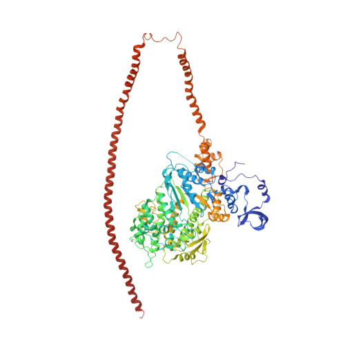
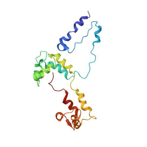
    3JAX

  • PubMed Abstract: 

    Muscle tissues are classically divided into two major types, depending on the presence or absence of striations. In striated muscles, the actin filaments are anchored at Z-lines and the myosin and actin filaments are in register, whereas in smooth muscles, the actin filaments are attached to dense bodies and the myosin and actin filaments are out of register. The structure of the filaments in smooth muscles is also different from that in striated muscles. Here we have studied the structure of myosin filaments from the smooth muscles of the human parasite Schistosoma mansoni. We find, surprisingly, that they are indistinguishable from those in an arthropod striated muscle. This structural similarity is supported by sequence comparison between the schistosome myosin II heavy chain and known striated muscle myosins. In contrast, the actin filaments of schistosomes are similar to those of smooth muscles, lacking troponin-dependent regulation. We conclude that schistosome muscles are hybrids, containing striated muscle-like myosin filaments and smooth muscle-like actin filaments in a smooth muscle architecture. This surprising finding has broad significance for understanding how muscles are built and how they evolved, and challenges the paradigm that smooth and striated muscles always have distinctly different components.


  • Organizational Affiliation

    Centro de Biología Estructural, Instituto Venezolano de Investigaciones Científicas (IVIC), Caracas 1020A, Venezuela; Department of Cell and Developmental Biology, University of Massachusetts Medical School, Worcester, MA 01655.


Macromolecules
Find similar proteins by:  (by identity cutoff)  |  3D Structure
Entity ID: 1
MoleculeChains Sequence LengthOrganismDetailsImage
myosin 2 heavy chain
A, B
974Schistosoma mansoniMutation(s): 0 
UniProt
Find proteins for A0A0R4I956 (Schistosoma mansoni)
Explore A0A0R4I956 
Go to UniProtKB:  A0A0R4I956
Entity Groups  
Sequence Clusters30% Identity50% Identity70% Identity90% Identity95% Identity100% Identity
UniProt GroupA0A0R4I956
Sequence Annotations
Expand
  • Reference Sequence
Find similar proteins by:  (by identity cutoff)  |  3D Structure
Entity ID: 2
MoleculeChains Sequence LengthOrganismDetailsImage
smooth muscle myosin essential light chain
C, D
151Schistosoma mansoniMutation(s): 0 
UniProt
Find proteins for A0A0R4I957 (Schistosoma mansoni)
Explore A0A0R4I957 
Go to UniProtKB:  A0A0R4I957
Entity Groups  
Sequence Clusters30% Identity50% Identity70% Identity90% Identity95% Identity100% Identity
UniProt GroupA0A0R4I957
Sequence Annotations
Expand
  • Reference Sequence
Find similar proteins by:  (by identity cutoff)  |  3D Structure
Entity ID: 3
MoleculeChains Sequence LengthOrganismDetailsImage
myosin regulatory light chain
E, F
196Schistosoma mansoniMutation(s): 0 
UniProt
Find proteins for A0A0R4I958 (Schistosoma mansoni)
Explore A0A0R4I958 
Go to UniProtKB:  A0A0R4I958
Entity Groups  
Sequence Clusters30% Identity50% Identity70% Identity90% Identity95% Identity100% Identity
UniProt GroupA0A0R4I958
Sequence Annotations
Expand
  • Reference Sequence
Experimental Data & Validation

Experimental Data

  • Method: ELECTRON MICROSCOPY
  • Resolution: 23.0 Å
  • Aggregation State: FILAMENT 
  • Reconstruction Method: HELICAL 
EM Software:
TaskSoftware PackageVersion
RECONSTRUCTIONEMAN2
RECONSTRUCTIONSPIDER

Structure Validation

View Full Validation Report


Entry History 

Deposition Data

Revision History  (Full details and data files)

  • Version 1.0: 2015-10-07
    Type: Initial release
  • Version 1.1: 2015-10-21
    Changes: Database references
  • Version 1.2: 2015-11-04
    Changes: Database references
  • Version 1.3: 2018-07-18
    Changes: Author supporting evidence, Data collection